
تصویر: Detailed 3D Illustration of Healthy Bone Structure and Density
منتشر شده:
آخرین به روز رسانی: ۲۲ مهٔ ۲۰۲۶ ساعت ۱۸:۰۳:۴۲ (UTC)
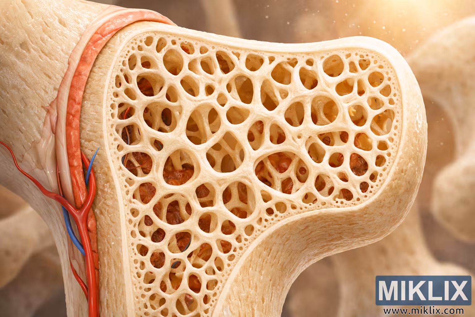
High-resolution 3D medical illustration of healthy bone structure and density, featuring cortical bone, periosteum, trabecular bone, and vascular bone marrow in a detailed cross-sectional view.
Detailed 3D Illustration of Healthy Bone Structure and Density

نسخههای موجود از این تصویر
فایلهای تصویری که در زیر برای دانلود موجود است، نسبت به تصاویر موجود در مقالات و صفحات این وبسایت که برای کاهش مصرف پهنای باند، از نظر حجم فایل بهینهتر شدهاند، فشردهسازی کمتری داشته و وضوح بالاتری دارند - و در نتیجه، کیفیت بالاتری دارند.
اندازه معمولی (1,536 x 1,024)
سایز بزرگ (3,072 x 2,048)
سایز بسیار بزرگ (4,608 x 3,072)
سایز فوق العاده بزرگ (6,144 x 4,096)
اندازهی کمیک بزرگ (1,048,576 x 699,051)
- هنوز در حال آپلود هستم... ;-)
شرح تصویر
This image presents a highly detailed, 3D-rendered medical illustration of healthy bone structure and density, designed to visually communicate the complex architecture of human bone tissue. The composition is in landscape orientation, with the bone shown in cross-section so that both the outer compact layer and the inner spongy region are clearly visible. The left side of the image is dominated by the cortical bone, which appears as a dense, smooth, and continuous off-white shell. Its surface has a subtle gloss, suggesting strength and solidity, and it curves gently, following the natural contour of a long bone. Surrounding this cortical layer is the periosteum, a thin but distinct pinkish membrane that hugs the outer surface of the bone. The periosteum is depicted as slightly translucent and richly vascularized, with fine blood vessels branching across it, emphasizing its role in nourishing and protecting the bone.
Emerging from the periosteum and penetrating into the cortical bone are clearly defined blood vessels. A prominent red vessel, representing an artery, and a smaller blue vessel, representing a vein, are shown entering through a small opening in the cortical layer. These vessels are rendered with smooth, tubular forms and subtle shading, making them appear three-dimensional and lifelike. Their presence highlights the dynamic, living nature of bone tissue, which is constantly supplied with nutrients and oxygen. The transition from the periosteum into the cortical bone is seamless yet visually distinct, with the vessels gently curving as they pass inward, reinforcing the sense of anatomical realism.
As the viewer’s eye moves from left to right, the illustration transitions from the dense cortical bone into the inner region of trabecular, or spongy, bone. This inner portion occupies the right side of the image and is depicted as an intricate lattice of creamy white struts known as trabeculae. These trabeculae form a highly interconnected, porous network, with numerous small cavities and channels between them. The structure is complex yet orderly, suggesting both lightness and strength. Each trabecula is carefully shaded to convey depth and curvature, and the overall pattern resembles a delicate, three-dimensional scaffold. The density and connectivity of the trabeculae visually communicate the idea of a healthy bone with robust structural integrity.
Within the spaces between the trabeculae lies the bone marrow, rendered in rich shades of red and deep crimson. This marrow appears soft and organic, contrasting with the firm, pale trabecular framework that surrounds it. Fine, branching blood vessels weave through the marrow, reinforcing its role as a vital, living tissue involved in blood cell production. The color contrast between the creamy white trabeculae and the reddish marrow creates a striking visual balance, making it easy to distinguish the different components while still feeling cohesive and natural. The interplay of light and shadow within these spaces adds depth and realism, drawing the viewer into the microscopic world inside the bone.
The lighting in the illustration is warm and inviting, with a gentle, glowing light source positioned toward the upper right corner of the scene. This light casts soft highlights on the cortical bone and trabeculae, emphasizing their textures and contours. Tiny particles of light appear to float in the air, especially near the glowing region, giving the image a subtle, almost atmospheric quality. These particles help create a sense of depth and three-dimensional space, as if the viewer is peering into a magnified, illuminated cross-section of living tissue. The warm tones of the light also enhance the natural colors of the bone and marrow, making the overall palette feel harmonious and lifelike.
In the background, beyond the main cross-section, there is a softly blurred suggestion of additional bones. These background elements are intentionally out of focus, rendered in muted beige and cream tones, so they do not compete with the primary subject. Instead, they provide context, hinting that the featured cross-section is part of a larger skeletal system. The blurred background also contributes to the depth of field, reinforcing the idea that the viewer’s attention is meant to remain on the detailed structure in the foreground. This selective focus mimics the effect of a macro lens in photography, where the main subject is sharply defined while the surroundings gently fade away.
The overall composition is carefully balanced, with the solid cortical bone on the left gradually giving way to the more open, lattice-like trabecular bone on the right. This left-to-right transition visually narrates the journey from the outer protective shell to the inner supportive framework and marrow-filled cavities. The cortical bone’s smooth, continuous surface conveys stability and protection, while the trabecular region’s intricate network suggests adaptability and efficient load distribution. Together, they illustrate how healthy bone achieves both strength and lightness through its hierarchical structure.
From an educational perspective, the illustration is designed to be both scientifically informative and visually engaging. The clear differentiation between cortical bone, periosteum, trabecular bone, and bone marrow makes it suitable for use in medical textbooks, patient education materials, or classroom presentations. The realistic rendering of blood vessels and the careful attention to color and texture help viewers understand that bone is not a static, inert material but a dynamic, living tissue. The dense, interconnected trabeculae and the rich, vascular marrow visually reinforce the concept of healthy bone density, making this image an effective tool for explaining conditions related to bone health, such as osteoporosis, in contrast to the robust structure shown here.
Overall, this high-resolution, landscape-oriented illustration offers a vivid, anatomically grounded view of healthy bone structure and density. The combination of detailed textures, realistic lighting, and thoughtful composition creates an image that is both aesthetically pleasing and scientifically accurate. It invites viewers to appreciate the complexity and elegance of bone architecture, highlighting how its layered design—from periosteum to cortical bone to trabecular network and marrow—works together to support, protect, and sustain the human body.
تصویر مربوط به: A Guide to the Health Benefits of Dandelions