
Image: Collagen Types and Their Functions
Published: June 24, 2025 at 10:42:57 AM UTC
Last updated: September 26, 2025 at 10:12:39 AM UTC
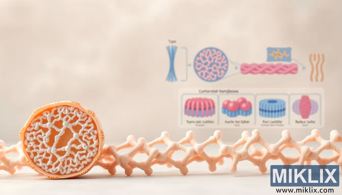
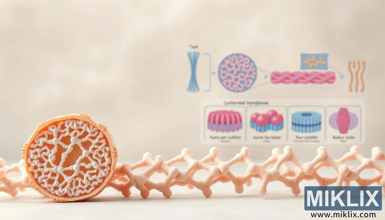
High-resolution, scientific illustration of collagen types I-V, highlighting structures, locations, and roles in the human body.

Available versions of this image
The image files available for download below are less compressed and higher resolution - and as a result of that, higher quality - than the images embedded in articles and pages on this website, which are more optimized for file size in order to reduce bandwidth consumption.
Regular size (1,344 x 768)
Large size (2,688 x 1,536)
Very large size (4,032 x 2,304)
Extra large size (5,376 x 3,072)
Comically large size (1,048,576 x 599,186)
- Still uploading... ;-)
Image description
The image offers a strikingly clear and scientifically grounded representation of collagen, the most abundant protein in the human body and a cornerstone of structural integrity within connective tissues. At the forefront, a magnified cross-sectional view of collagen fibrils captures the complexity of their architecture, revealing the intricate lattice-like arrangement that provides both tensile strength and flexibility. The pattern is rendered with photorealistic precision, allowing the viewer to perceive the depth and interwoven nature of these molecular structures. This detailed portrayal emphasizes the crucial role of collagen fibrils as the scaffolding upon which tissues like skin, cartilage, bone, and tendons are built. The texture, color contrast, and three-dimensional appearance of the fibril highlight not only its biological function but also its aesthetic beauty, turning a microscopic structure into a compelling visual centerpiece.
Moving into the middle ground, schematic diagrams are incorporated to contextualize the molecular imagery by linking it to the biological roles of different collagen types. Each diagram illustrates the functional specialization of the main collagen families: type I collagen, depicted as densely packed and strong, is associated with skin, tendons, and bones, where it provides tensile strength and durability; type II collagen is shown in relation to cartilage, highlighting its role in cushioning joints and maintaining mobility; type III collagen, often paired with type I, supports the pliability of organs, skin, and vascular tissues; type IV collagen is represented in basement membranes, where its sheet-like structure forms filtration barriers and underpins cellular attachment; and type V collagen is portrayed as a regulator of fibril assembly, essential in maintaining the proper diameter and organization of other collagen fibers. The clear, color-coded schematic design ensures that the viewer can easily distinguish these collagen subtypes while appreciating their unique contributions to the body’s structural harmony.
The background provides an understated yet essential layer to the composition. A soft, muted palette of warm neutrals and delicate organic gradients creates an atmosphere that feels both clinical and approachable, allowing the scientific content to stand out without overwhelming the senses. This subtle backdrop mirrors the environment of an educational or research setting, lending the image an academic credibility while also making it visually soothing. It evokes the impression of being within a laboratory or anatomical atlas, where clarity and precision are paramount, yet the design still retains a touch of artistic elegance.
Lighting plays a crucial role in bringing the illustration to life. Gentle highlights and shadows define the three-dimensional form of the collagen fibrils, giving them volume and tangibility, while the schematic diagrams are evenly illuminated to maintain readability and accuracy. The interplay of photorealistic textures in the foreground with clean, diagrammatic lines in the middle ground creates a seamless fusion between artistic visualization and scientific instruction. This duality ensures that the image appeals equally to academic audiences seeking precision and to broader viewers curious about the invisible structures that sustain human life.
Altogether, the composition succeeds in transforming a subject as microscopic and abstract as collagen proteins into a vivid and comprehensible visual narrative. It bridges molecular science and human physiology, illustrating how something as small as a protein fiber governs essential aspects of strength, elasticity, and resilience in the body. By highlighting both the intricate molecular design and the macroscopic functions of collagen types I through V, the image conveys not only factual knowledge but also a sense of wonder at the sophistication of human biology. It reminds the viewer that beneath the surface of skin and tissue lies a world of organized complexity, one that sustains movement, protection, and vitality throughout life.
The image is related to: From Skin to Joints: How Daily Collagen Gives Your Whole Body a Boost