
Image: Beta Alanine Molecular Mechanism
Published: June 23, 2025 at 3:07:04 PM UTC
Last updated: September 26, 2025 at 10:09:15 AM UTC
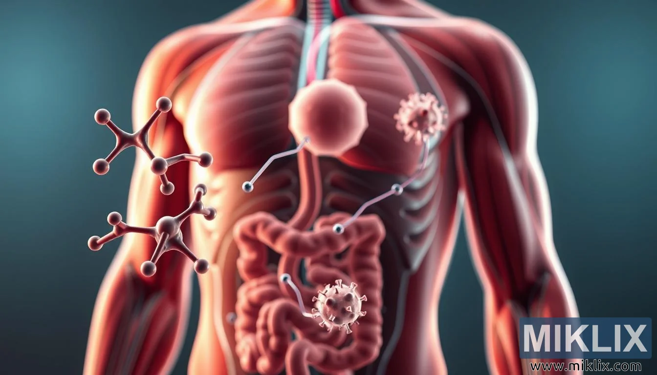
Detailed 3D illustration showing beta alanine absorption, muscle carnosine formation, and lactic acid buffering for performance.

Available versions of this image
The image files available for download below are less compressed and higher resolution - and as a result of that, higher quality - than the images embedded in articles and pages on this website, which are more optimized for file size in order to reduce bandwidth consumption.
Regular size (1,344 x 768)
Large size (2,688 x 1,536)
Very large size (4,032 x 2,304)
Extra large size (5,376 x 3,072)
Comically large size (1,048,576 x 599,186)
- Still uploading... ;-)
Image description
The image presents a scientifically rich and visually precise 3D render of the human body’s response to beta alanine supplementation, designed to illustrate both molecular and physiological processes in a way that is both accessible and authoritative. At first glance, the focus falls on the central figure of a semi-transparent human torso, where muscle fibers, vascular pathways, and digestive structures are revealed in a clinical cutaway style. This anatomical accuracy provides the framework upon which the molecular journey of beta alanine is mapped, transforming abstract biochemical processes into a compelling visual narrative.
In the foreground, stylized molecular models of beta alanine are shown as interconnected spheres, their simple yet elegant geometry signifying the building blocks of the compound. These molecules are depicted entering the digestive system, absorbed through the intestinal walls into the bloodstream. Their presence here highlights the initial step in the supplementation process—how something ingested transforms into a circulating agent capable of affecting performance at the muscular level. The clarity with which the molecules are presented underscores the scientific intent of the rendering: to demystify the unseen and make the molecular mechanics of supplementation tangible.
As the eye moves toward the middle ground, the focus shifts to muscle tissue. Arterial pathways are traced visually as conduits transporting beta alanine molecules directly into muscle cells, where they encounter histidine. The rendering demonstrates this molecular union with precision, depicting beta alanine and histidine combining to form carnosine. This moment, though microscopic, is represented on a scale that allows viewers to appreciate its importance. By zooming into this critical interaction, the image effectively communicates the biochemical transformation at the heart of beta alanine’s impact on athletic performance.
In the background, the broader physiological outcome is revealed: elevated levels of carnosine within muscle fibers. This increase is symbolized by glowing molecular clusters embedded within the muscle tissue, visually representing heightened buffering capacity. The scene illustrates how carnosine counteracts lactic acid buildup, delaying the onset of fatigue and enabling improved endurance. The muted tones and controlled lighting used in this part of the composition lend it a clinical authority, ensuring that the scientific message is conveyed with both precision and clarity.
What makes the render particularly compelling is the way it bridges multiple scales of biology—molecular, cellular, and systemic—within a single frame. By transitioning smoothly from the digestive tract to the bloodstream, then to the microscopic environment of muscle cells, and finally to the macroscopic effect on the entire musculature, the image creates a holistic view of beta alanine’s role. The shallow depth of field subtly directs the viewer’s attention, ensuring focus rests on the processes most critical to understanding the supplement’s function.
Altogether, the composition conveys more than just anatomy and molecules—it tells a story of transformation, from ingestion to performance enhancement. The use of restrained colors and sharp lighting balances clarity with clinical realism, avoiding distraction while reinforcing the scientific tone. The result is a powerful educational visualization that captures both the complexity and the elegance of beta alanine’s role in human physiology, making it an indispensable tool for conveying the supplement’s mechanism of action to athletes, students, and medical professionals alike.
The image is related to: The Carnosine Catalyst: Unlocking Muscle Performance with Beta-Alanine