
이미지: Laboratory Analysis of Alpha Acid Crystal Formation
게시됨:
마지막으로 업데이트되었습니다: 2026년 9월 20일 오전 9시 57분 40초 UTC
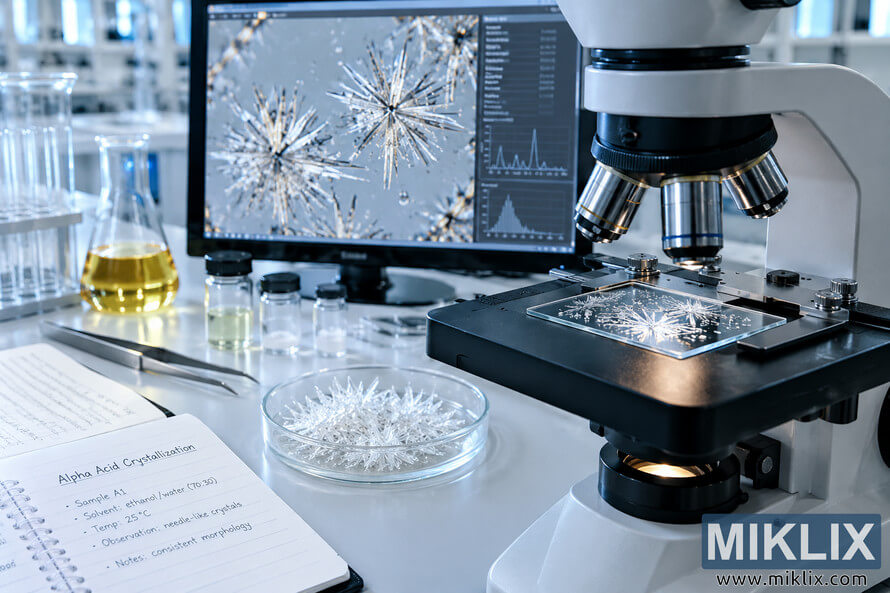
High-resolution laboratory scene showing microscopic analysis of alpha acid crystal formation, with needle-like crystals, scientific equipment, samples, glassware, experimental notes, and digital analysis.
Laboratory Analysis of Alpha Acid Crystal Formation
이 이미지의 사용 가능한 버전
아래에서 다운로드할 수 있는 이미지 파일은 이 웹사이트의 기사 및 페이지에 삽입된 이미지보다 압축률이 낮고 해상도가 높으며, 대역폭 소비를 줄이기 위해 파일 크기에 더 최적화되어 있어 품질이 더 높습니다.
일반 사이즈 (1,536 x 1,024)
대형 사이즈 (3,072 x 2,048)
매우 큰 크기 (4,608 x 3,072)
초대형 사이즈 (6,144 x 4,096)
코믹하게 큰 사이즈 (1,048,576 x 699,051)
- 아직 업로드 중... ;-)
이미지 설명
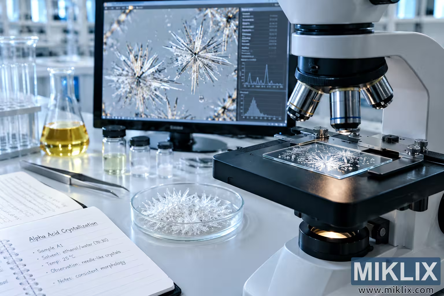
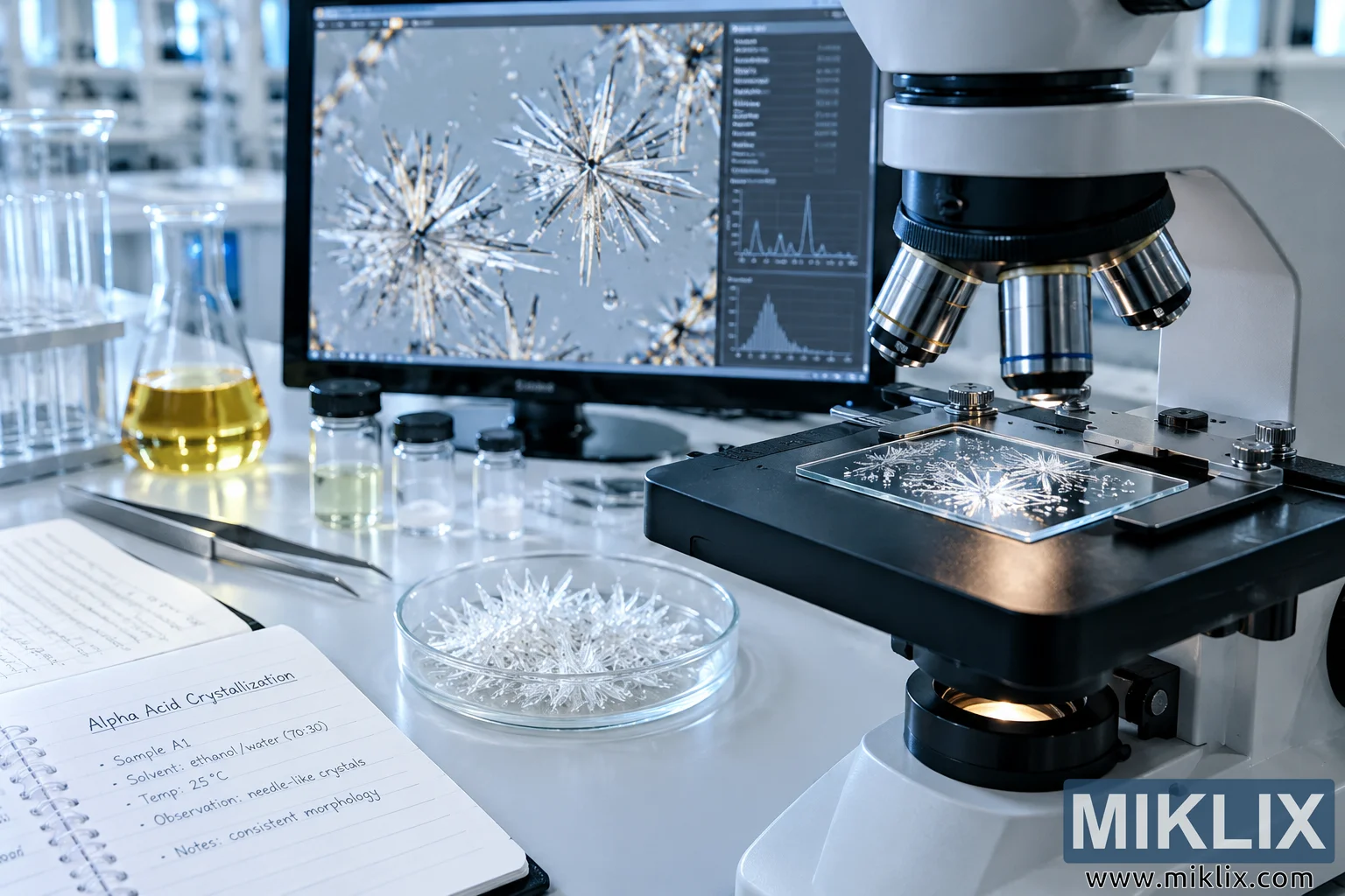
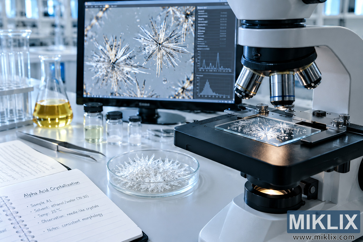
A high-resolution landscape photograph depicts a clean, modern laboratory workspace focused on the microscopic analysis of alpha acid crystal formation. The composition has the polished realism of professional scientific and technical photography, with crisp detail across the primary laboratory equipment and a softly receding background. On the right side of the image, a large compound microscope dominates the foreground. Its pale gray and black body is shown in close detail, including the rotating objective turret, several metallic objective lenses, the mechanical stage, adjustment controls, and the illuminated base. The microscope is positioned at a slight angle, allowing the viewer to see both the instrument itself and the sample being examined. A rectangular glass microscope slide rests on the dark stage beneath the objective lenses. Across the slide are numerous bright, branching crystalline structures. Their elongated, needle-like forms radiate outward into intricate clusters, producing patterns reminiscent of frost, delicate mineral growth, or microscopic starbursts. The intense illumination from below makes the crystalline material appear almost white and slightly translucent against the darker surface of the microscope stage.
Behind the microscope, a large laboratory monitor occupies much of the central and upper-left portion of the scene. The screen displays a magnified microscopy image of similar crystal formations, reinforcing the impression that the sample on the microscope is being actively analyzed. Several prominent crystals form radial clusters made from long, slender needles. Smaller particles and droplets are scattered around the larger structures. Along the right side of the monitor interface are scientific analysis panels containing rows of measurement data and multiple graphical displays. These include a line graph with a pronounced peak and another distribution-style graph below it. The interface is dark and technical in appearance, contrasting with the bright microscopy image and the white laboratory surroundings. No recognizable commercial branding is intentionally featured as part of the scene.
In the center foreground, directly in front of the monitor and beside the microscope, sits a clear glass petri dish containing a dense collection of pale crystalline material. The crystals have sharp, elongated, branching shapes similar to those visible on the microscope slide and monitor. Light passing through the glass and crystal structures creates bright reflections and highlights, emphasizing their delicate geometry. The petri dish serves as a larger-scale visual representation of the material being studied and helps connect the physical sample preparation process with the microscopic analysis taking place nearby.
To the left of the petri dish are several small transparent laboratory sample vials with dark caps. Some contain clear or faintly tinted liquid, while others appear nearly empty or contain small sample quantities. Behind these containers is an Erlenmeyer flask holding a transparent golden-yellow liquid. The liquid catches the cool laboratory lighting and provides a warm visual contrast to the otherwise white, silver, gray, and blue-toned environment. Additional glassware and a rack of test tubes can be seen farther to the left and in the softly blurred background, establishing the setting as an active analytical laboratory rather than an isolated product shot.
A pair of long metallic laboratory tweezers or forceps lies diagonally on the work surface near the sample containers. Their placement suggests recent or ongoing sample handling. The polished metal reflects the bright overhead lighting and contributes to the precise, sterile character of the workspace. The laboratory bench itself is smooth, clean, and light-colored, with enough subtle reflection to enhance the impression of a carefully maintained scientific environment.
In the lower-left foreground, an open spiral-bound laboratory notebook provides contextual information about the experiment. The visible page is headed "Alpha Acid Crystallization" and contains handwritten-style notes describing a sample, solvent conditions, temperature, observations, and crystal morphology. The notes indicate an experimental workflow centered on crystallization and microscopic examination. The notebook is positioned naturally beside the instruments rather than presented as a formal graphic, giving the scene the feeling of a real laboratory investigation in progress. Its presence also helps communicate that the image represents documentation and analysis rather than simply decorative crystal imagery.
The lighting is bright, cool, and clinical, with strong but controlled highlights on glass, polished metal, microscope components, and crystalline structures. A shallow-to-moderate depth of field keeps the microscope, slide, petri dish, notebook, and monitor clearly recognizable while allowing distant laboratory equipment to blur gently into the background. The overall palette consists primarily of clean whites, cool grays, metallic silver, black instrument components, subtle blue tones, and the warm yellow of the liquid in the flask. This restrained color scheme reinforces the scientific atmosphere while allowing the luminous crystal formations to become the central visual subject.
The image communicates a laboratory investigation of alpha acid crystallization through several interconnected visual cues: a physical crystalline sample, a prepared microscope slide, magnified crystal imagery on a computer display, analytical graphs, labeled experimental notes, chemical glassware, and precision laboratory instruments. The crystal formations themselves are the strongest recurring motif. Their fine, needle-like structures and radial branching patterns appear at multiple scales throughout the composition, visually illustrating crystal growth and morphology. The microscope anchors the scientific process, while the monitor suggests digital imaging and quantitative analysis. Together, these elements create a convincing representation of analytical chemistry or materials research in which crystallization behavior is being observed, recorded, and evaluated under controlled laboratory conditions. The photograph is presented in a wide landscape format, making it suitable for scientific editorial material, laboratory research content, technical articles, educational pages, or visual communication concerning alpha acid crystal formation and microscopic chemical analysis.
이 이미지는 다음과 관련이 있습니다: Hops in Beer Brewing: Columbus