
Afbeelding: Corn Smut Infection on Maize Ear
Gepubliceerd:
Laatst bijgewerkt: 13 maart 2026 om 20:38:32 UTC
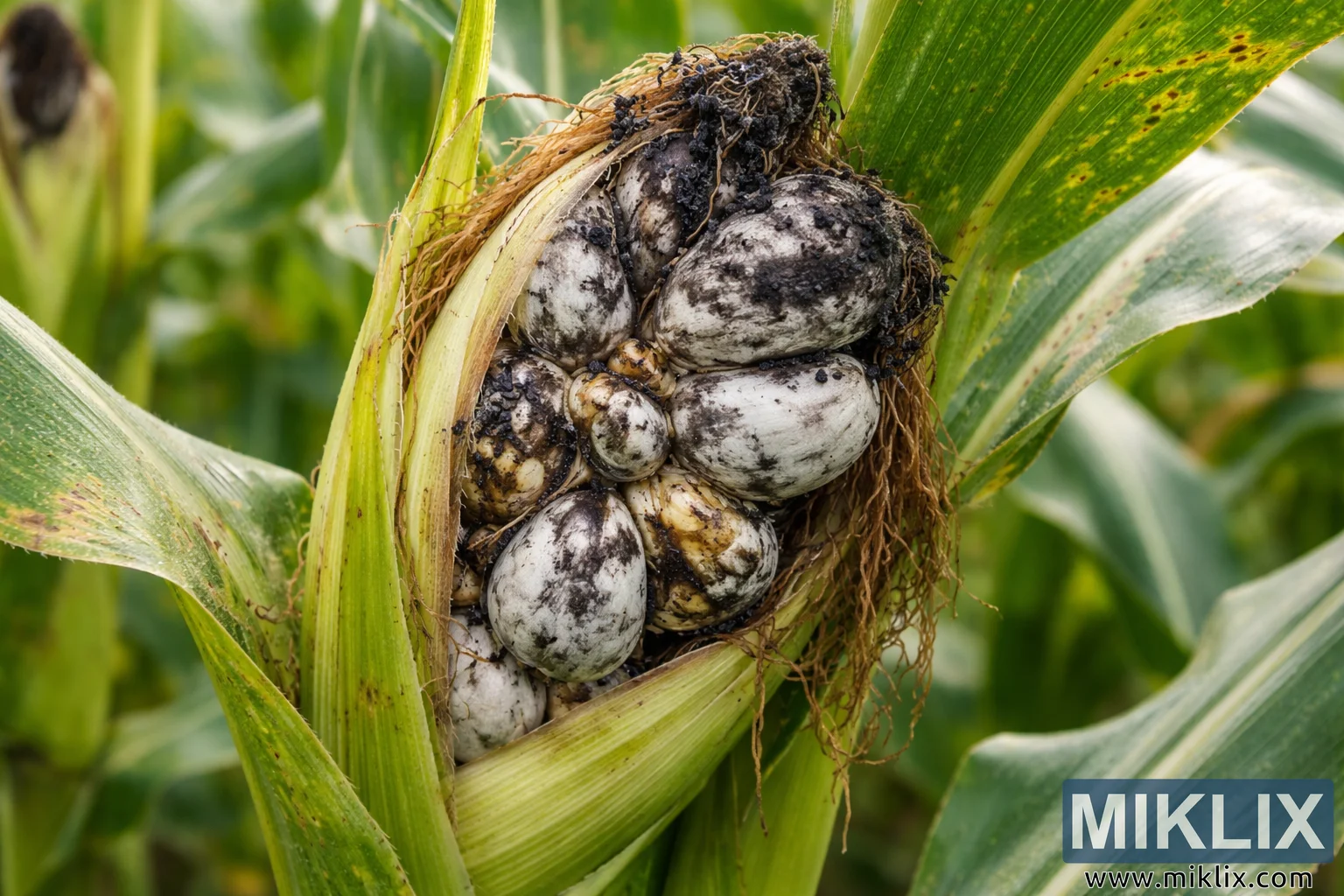
High-resolution close-up image of a corn plant infected with corn smut (Ustilago maydis), displaying swollen gray galls and black spores replacing the kernels on the maize ear.
Corn Smut Infection on Maize Ear

Beschikbare versies van deze afbeelding
De afbeeldingsbestanden die hieronder kunnen worden gedownload, zijn minder gecomprimeerd en hebben een hogere resolutie - en daardoor een hogere kwaliteit - dan de afbeeldingen die zijn ingesloten in artikelen en pagina's op deze website, die meer zijn geoptimaliseerd voor bestandsgrootte om het bandbreedtegebruik te beperken.
Normale maat (1,536 x 1,024)
Groot formaat (3,072 x 2,048)
Zeer groot formaat (4,608 x 3,072)
Extra groot formaat (6,144 x 4,096)
Komisch groot formaat (1,048,576 x 699,051)
- Nog steeds aan het uploaden... ;-)
Beschrijving afbeelding
This high-resolution landscape photograph shows a close-up view of a corn plant visibly infected with corn smut, a fungal disease caused by Ustilago maydis. The focal point of the image is a partially opened corn ear where the normal rows of yellow kernels have been replaced by swollen, irregularly shaped fungal galls. These bulbous structures appear grayish-white to pale silver in color and vary in size, forming a clustered mass that fills the interior of the husk. Several galls display dark, mottled surfaces where the outer membrane has begun to rupture, exposing the black, powdery spores characteristic of the disease.
The husk leaves surrounding the ear are pulled back slightly, revealing the infected tissue. They appear light green with subtle striations and natural curvature, framing the diseased ear and guiding the viewer's attention toward the center of the image. Fine strands of corn silk emerge from the top of the ear, tangled among the fungal growths. The silk fibers are dry and brown, suggesting a later stage of development in the corn plant as well as the progression of the infection.
Across the galls, dark speckles and patches of spore masses create a striking contrast against the lighter gray outer membranes. Some galls are smooth and intact, while others show cracking or fragmentation where spores have begun to disperse. This variation illustrates different stages of the fungal lifecycle occurring simultaneously within the same ear.
The background is softly blurred with shallow depth of field, showing green corn leaves and stalks that suggest a larger maize field environment. This natural backdrop provides contextual information while ensuring that the infected ear remains the clear subject of the photograph. The lighting appears natural and even, likely captured outdoors during daylight, allowing the textures of the plant tissue and fungal structures to be clearly visible.
Fine details such as the fibrous strands of silk, the subtle veining of the husk leaves, and the granular appearance of the black spores contribute to the scientific and documentary quality of the image. The photograph effectively highlights the dramatic morphological changes caused by corn smut infection, transforming what would normally be a uniform ear of corn into a cluster of swollen fungal growths. As a result, the image serves both as a visually striking natural scene and as an educational depiction of a well-known plant disease affecting maize crops.
De afbeelding is gerelateerd aan: Growing Corn: Your Complete Guide to Sweet Success in the Garden