
Obrázok: Microscopic Battle: Immune Cells Fighting Pathogens
Publikované:
Posledná aktualizácia: 22. mája 2026 o 18:03:28 UTC
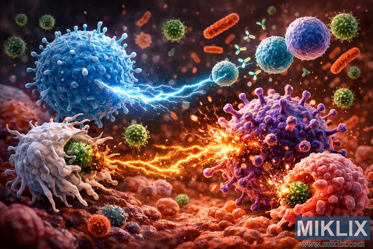
Highly detailed microscopic illustration of immune cells fighting pathogens, featuring T cells, B cells, macrophages, natural killer cells, and neutrophils attacking viruses and bacteria in a dramatic, glowing biological environment.
Microscopic Battle: Immune Cells Fighting Pathogens

Dostupné verzie tohto obrázka
Obrázkové súbory, ktoré sú k dispozícii na stiahnutie nižšie, sú menej komprimované a majú vyššie rozlíšenie - a vďaka tomu aj vyššiu kvalitu - ako obrázky vložené do článkov a stránok na tejto webovej lokalite, ktoré sú viac optimalizované z hľadiska veľkosti súborov, aby sa znížila spotreba šírky pásma.
Bežná veľkosť (1,536 x 1,024)
Veľká veľkosť (3,072 x 2,048)
Veľmi veľká veľkosť (4,608 x 3,072)
Extra veľká veľkosť (6,144 x 4,096)
Komicky veľká veľkosť (1,048,576 x 699,051)
- Stále nahrávam... ;-)
Popis obrázku
A highly detailed, digitally rendered microscopic scene depicts a dramatic battle between the human immune system and invading pathogens, visualized as if the viewer were shrunk down to the cellular scale. The composition is set in a warm, reddish biological environment that suggests tissue or blood, softly blurred in the background to keep the focus on the intense interactions between immune cells and microbes. Countless tiny glowing particles, fragments of debris, and suspended specks of light drift through the scene, reinforcing the sense of a dynamic, living battlefield.
On the center-left of the image, a large, spherical blue T cell dominates the frame. Its surface is textured and slightly rough, covered with numerous short, spike-like protrusions that give it a distinctive, almost armored appearance. From the front of this T cell, a concentrated stream of glowing, electric-blue energy bursts outward, arcing across the space like miniature lightning. This energy beam is directed toward a pathogen on the right side of the scene, visually representing the T cell’s targeted attack on infected or foreign cells. The glow from this energy illuminates nearby particles and casts a cool blue reflection on the surrounding environment, contrasting sharply with the warmer tones of the pathogens.
To the left of the T cell, a white macrophage appears as a large, rounded cell with a soft, almost gelatinous surface. Its membrane is extended into multiple tentacle-like pseudopodia that reach outward in fluid, organic curves. These pseudopodia are wrapped around a green, spiky bacterium, whose rigid, thorn-like projections contrast with the macrophage’s flexible tendrils. The bacterium appears to be in the process of being engulfed: parts of its surface are dissolving, breaking into tiny fragments and glowing particles as it is pulled inward. The macrophage’s pseudopodia form a partial cage around the bacterium, visually communicating the process of phagocytosis, where the immune cell literally consumes the invader.
In the lower central area of the illustration, an orange natural killer (NK) cell adds another focal point. This NK cell is roughly spherical but covered in short, uneven spikes and ridges, giving it a rugged, aggressive texture. Its coloration ranges from deep orange to fiery yellow highlights, suggesting heat and intensity. From its surface, several bright, glowing tendrils of energy extend outward like flaming whips. These tendrils lash toward a large purple virus particle on the right side of the image. The virus is spherical, studded with numerous spike-like proteins that protrude evenly from its surface, giving it a crown-like silhouette. Where the NK cell’s energy tendrils strike the virus, a small explosion of light and debris erupts—tiny fragments of the viral shell and glowing particles scatter outward, indicating that the virus is being damaged or destroyed.
Scattered around the scene are smaller, round blue B cells. These cells are smoother and more compact than the T cell, with subtle surface texture but fewer protrusions. From the B cells, streams of glowing green Y-shaped antibodies shoot outward in multiple directions. These antibodies travel through the space like tiny guided projectiles, some targeting the large purple virus particle, others heading toward rod-shaped bacteria in the upper right region of the image. The antibodies are depicted with crisp, recognizable Y shapes, making their identity clear even at a small scale. Their bright green glow stands out against the reddish background and the cooler blues of the immune cells, visually emphasizing their role as precise molecular weapons.
In the background, several reddish-orange rod-shaped bacteria drift through the environment. They are elongated, with slightly rounded ends and subtle surface textures that suggest cell walls. Some appear intact, while others show signs of damage, with small cracks, missing fragments, or faint glows where antibodies or immune cell attacks have struck them. Interspersed among these rods are smaller green virus particles, each with a spherical body and short spikes, echoing the larger purple virus but on a smaller scale. These background pathogens add depth and context, suggesting that the battle extends beyond the immediate foreground.
On the lower right side of the illustration, a large reddish-pink neutrophil is engaged in another act of defense. The neutrophil has an irregular, somewhat lobed shape, with a soft, semi-translucent membrane that bulges outward in places. It is in the process of engulfing a smaller green, spiky bacterium, similar in appearance to the one being consumed by the macrophage. The bacterium is partially surrounded by the neutrophil’s membrane, which curves around it like a wave. Around the point of contact, glowing particles and faint streams of light represent digestive enzymes being released to break down the pathogen. The bacterium’s spikes appear to be dissolving, and tiny fragments float away, reinforcing the sense of active destruction.
Throughout the entire scene, lighting plays a crucial role in conveying drama and motion. Strong highlights and glows emanate from the points of interaction: the blue energy beam from the T cell, the fiery tendrils from the NK cell, the green antibodies from the B cells, and the enzyme-rich region around the neutrophil. These light sources cast subtle reflections and color shifts on nearby cells and particles, creating a rich interplay of cool blues, warm reds, oranges, and purples. The background remains softly blurred and predominantly reddish, suggesting a tissue or blood environment without drawing attention away from the central action.
The overall composition balances scientific inspiration with artistic interpretation. Each immune cell type—T cell, B cell, macrophage, NK cell, and neutrophil—is visually distinct in color, texture, and behavior, making it easy to differentiate their roles in the immune response. The pathogens, including the large purple virus particle, smaller green viruses, and rod-shaped bacteria, are equally stylized yet clearly recognizable as foreign invaders. The image captures a frozen moment in an ongoing microscopic war, emphasizing both the complexity and the power of the immune system as it identifies, targets, and neutralizes threats within the body.
Obrázok súvisí s: A Guide to the Health Benefits of Dandelions