
Slika: Detailed Cross-Section of the Human Eye Focusing on the Macula
Objavljeno:
Nazadnje posodobljeno: 3. september 2026 ob 8:25:56 pop. UTC
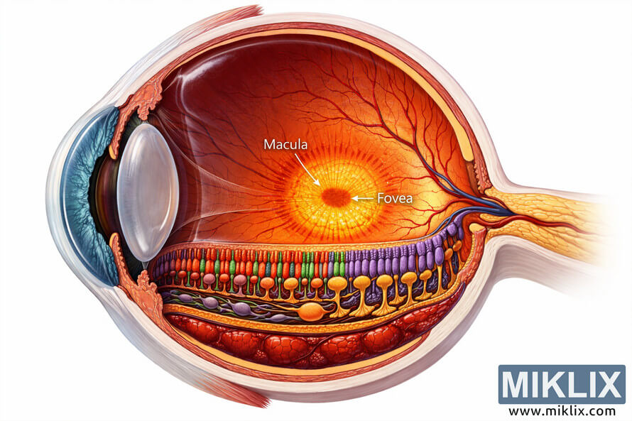
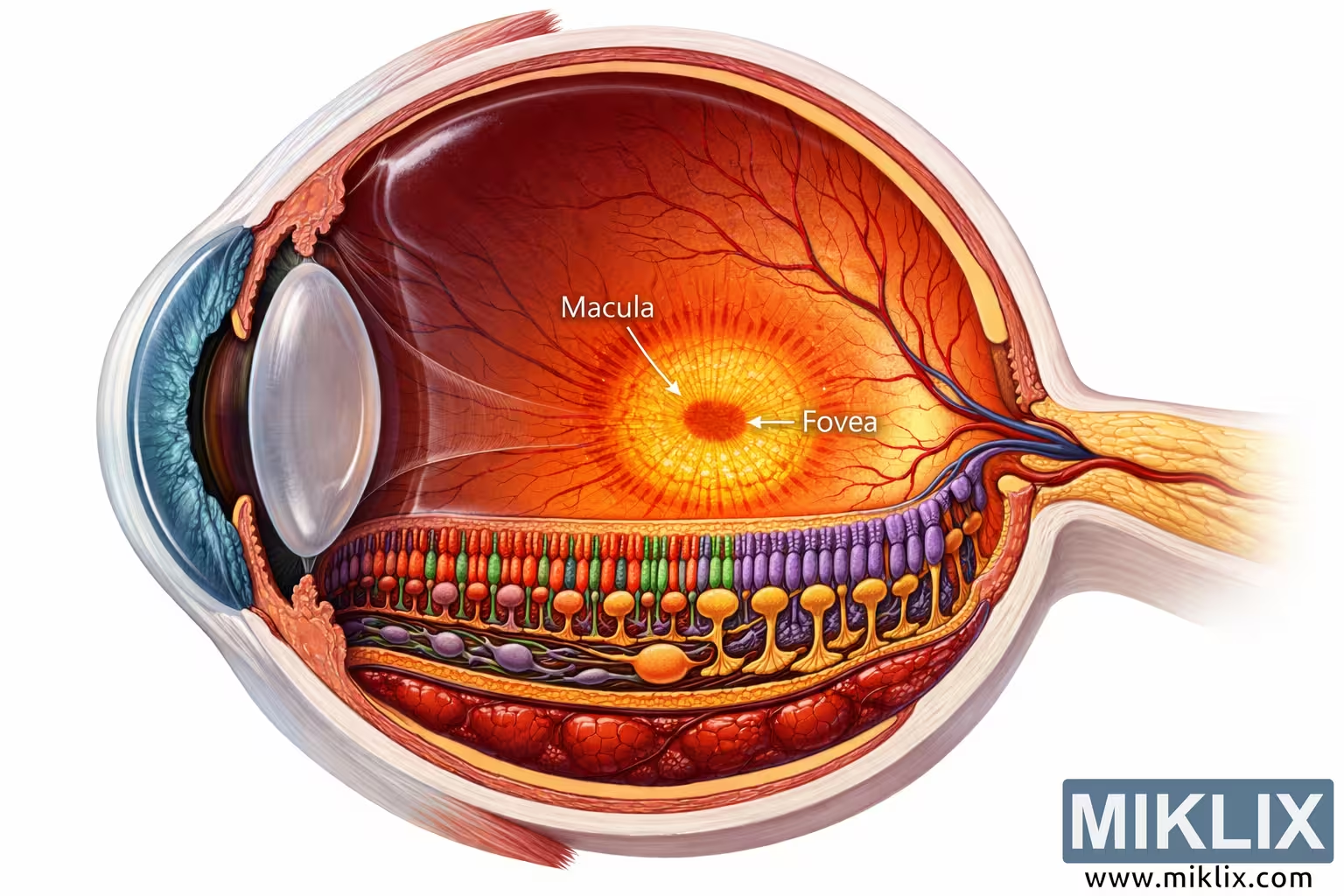
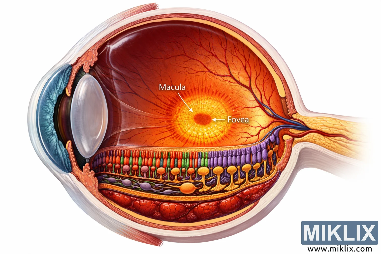
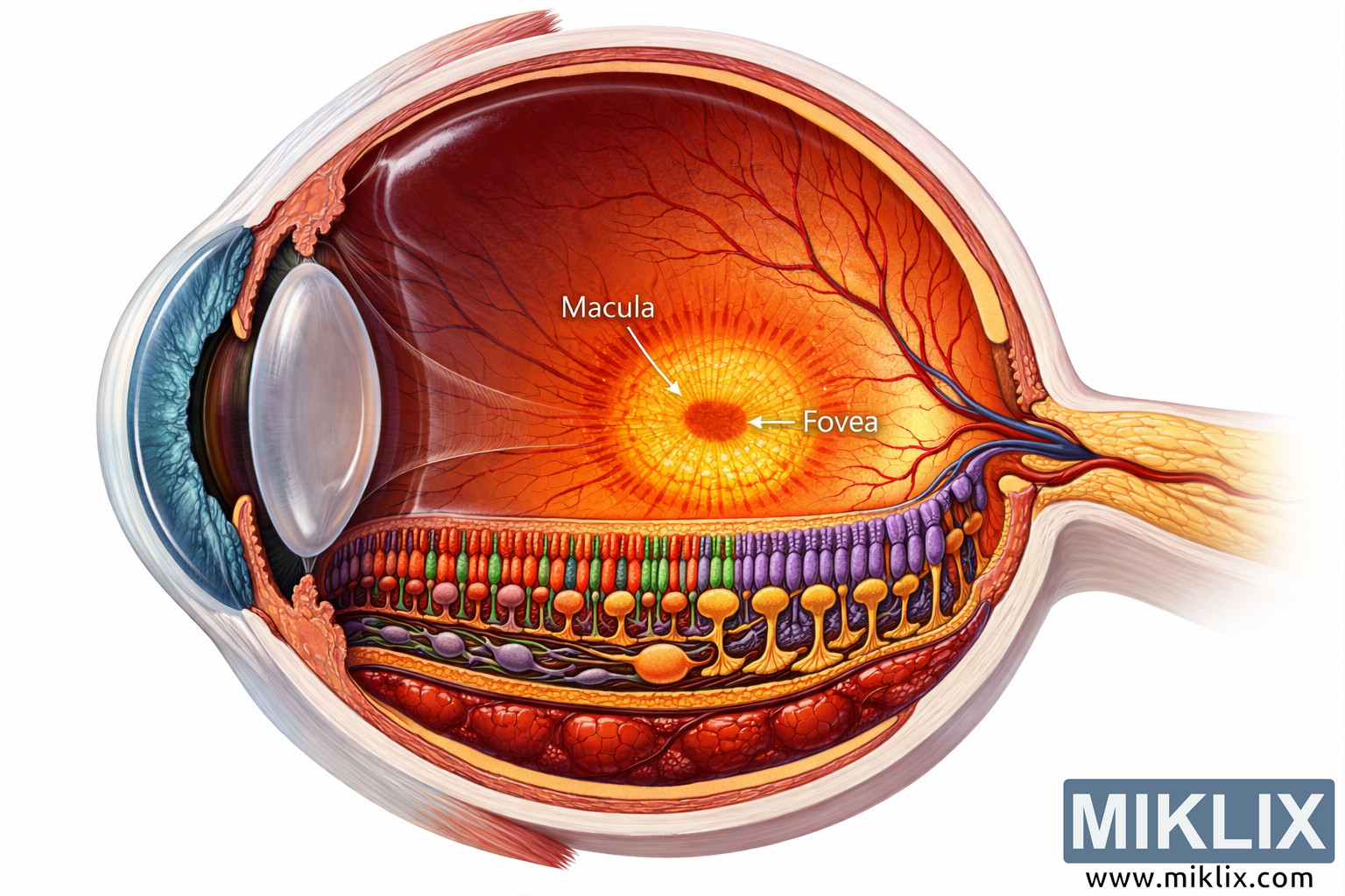
A high-resolution anatomical illustration of the human eye in cross-section, highlighting the macula and fovea. The image depicts the cornea, lens, retina, choroid, and optic nerve in vivid detail, offering an educational view of ocular anatomy and visual processing.
Detailed Cross-Section of the Human Eye Focusing on the Macula
Razpoložljive različice te slike
Slikovne datoteke, ki so na voljo za prenos spodaj, so manj stisnjene in imajo višjo ločljivost - in s tem višjo kakovost - kot slike, vdelane v članke in strani na tem spletnem mestu, ki so bolj optimizirane glede velikosti datotek, da se zmanjša poraba pasovne širine.
Redna velikost (1,536 x 1,024)
Velika velikost (3,072 x 2,048)
Zelo velika velikost (4,608 x 3,072)
Zelo velika velikost (6,144 x 4,096)
Komično velika velikost (1,048,576 x 699,051)
- Še vedno nalagam... ;-)
Opis slike
This image presents a meticulously detailed, high-resolution cross-sectional illustration of the human eye, emphasizing the macula—the region responsible for central, high-acuity vision. The composition is rendered in landscape orientation, allowing a comprehensive view of the eye’s internal anatomy from the cornea at the front to the optic nerve at the rear. The outermost layer, the sclera, forms a protective white shell around the eye, while the transparent cornea curves outward at the front, permitting light to enter. Behind the cornea lies the anterior chamber, filled with aqueous humor, a clear fluid that maintains intraocular pressure and nourishes the corneal and lens tissues. The iris, a circular band of muscle tissue, surrounds the pupil and regulates the amount of light entering the eye. The pupil appears as a dark central aperture, dynamically adjusting its diameter in response to light intensity.
Immediately behind the pupil is the crystalline lens, depicted as a transparent, biconvex structure suspended by fine zonular fibers. The lens focuses incoming light onto the retina, and its curvature changes to accommodate near and distant vision. The vitreous body occupies the large central cavity of the eye, filled with vitreous humor—a gel-like substance that maintains the spherical shape and optical clarity of the eye. Surrounding the vitreous chamber, the retina lines the inner surface, shown here as a multi-layered structure with intricate cellular organization.
The retina’s outermost layer, the retinal pigment epithelium (RPE), is illustrated as a dark brown band that absorbs stray light and supports photoreceptor metabolism. Above it, the photoreceptor layer contains rods and cones—the sensory cells responsible for converting light into neural signals. The cones, concentrated in the macula and especially in the fovea, are shown as short, tapered cells in shades of red, orange, and green, representing their sensitivity to different wavelengths of light. The rods, more numerous in peripheral regions, appear as elongated purple cells specialized for low-light vision. These photoreceptors connect to bipolar cells, depicted as slender green and yellow neurons that relay signals to ganglion cells. The ganglion cells, larger and orange in color, form the innermost retinal layer, their axons converging to create the optic nerve, which transmits visual information to the brain.
The macula, located near the center of the retina, is highlighted in warm tones of yellow and orange, signifying its dense concentration of cones and its role in fine detail perception. Within the macula lies the fovea, a small depression marked by an even higher density of cones and the absence of rods. This region is the focal point of sharp vision, enabling activities such as reading and recognizing faces. The illustration labels both the macula and fovea clearly, with arrows pointing to their respective positions. The surrounding retinal layers are shown with precise gradations of color and texture, emphasizing the complexity of neural pathways involved in vision.
Beneath the retina lies the choroid, a vascular layer rendered in deep red hues, representing its rich blood supply that nourishes the outer retinal layers. The choroid’s capillaries and connective tissue are subtly shaded to convey depth and biological realism. The sclera beneath the choroid appears as a thick, white fibrous layer, providing structural integrity and protection. On the far right, the optic nerve emerges as a thick bundle of pale yellow fibers extending toward the brain, symbolizing the transmission of visual signals.
Lighting throughout the illustration is even and balanced, ensuring clarity across all anatomical features. The color palette combines realistic tones with educational contrast—warm yellows and oranges for the macula, cool blues and purples for the anterior structures, and deep reds for vascular tissues. The overall composition achieves both scientific accuracy and aesthetic appeal, making it suitable for medical education, ophthalmology reference, and visual science communication.
The image’s focal emphasis on the macula and fovea underscores their importance in human vision. The surrounding structures—cornea, lens, vitreous body, retina, choroid, and optic nerve—are arranged harmoniously to illustrate the eye’s optical pathway. This depiction captures the delicate interplay between light transmission, photoreceptor activation, and neural processing, offering viewers a vivid understanding of how the eye converts light into sight. The result is a comprehensive visual narrative of ocular anatomy, rendered with precision, clarity, and depth.
Slika je povezana z: Loquat: The Sweet Fruit Packed with Real Health Benefits