
Image: Macro Close-Up of Rakau Hop Cones Revealing Lupulin Glands
Published: October 24, 2026 at 12:23:02 AM UTC
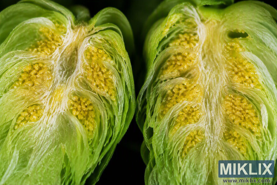
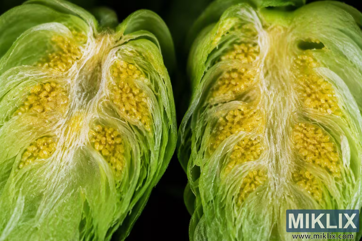
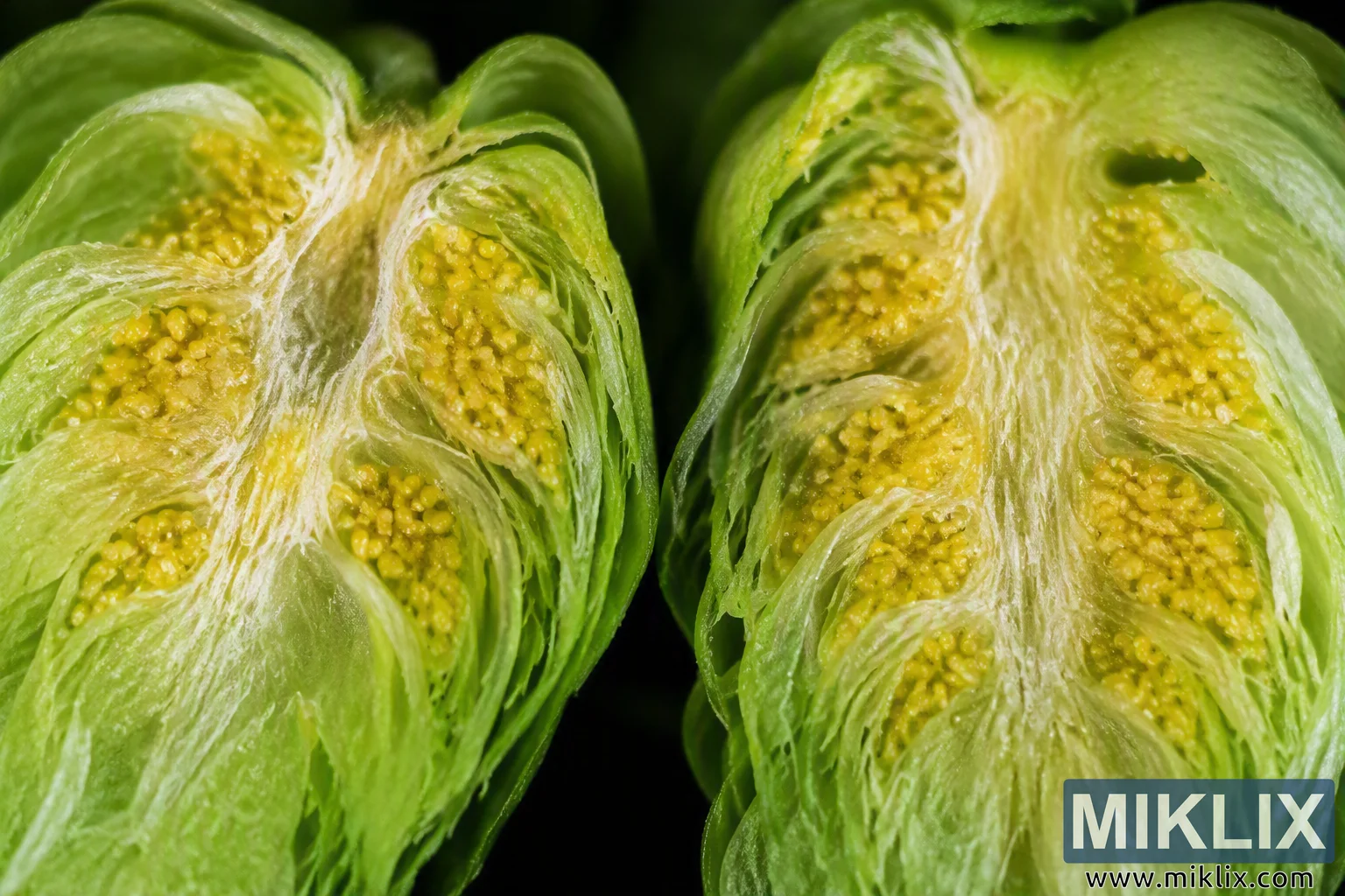
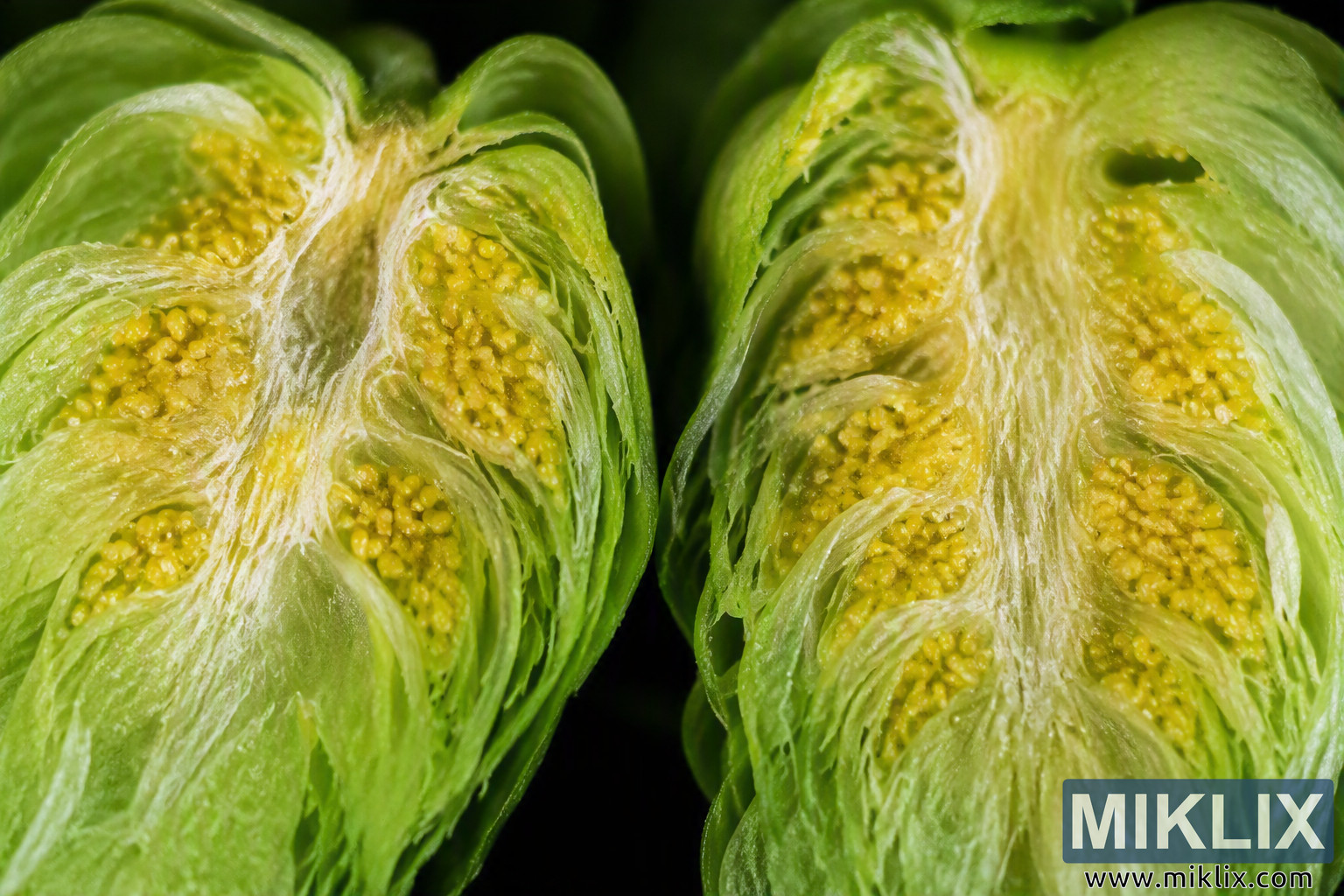
Detailed macro image of Rakau hop cones cut open to reveal vivid yellow lupulin glands nestled within layered green bracts. The close-up botanical photograph highlights texture, plant structure, and resin-rich hop anatomy.
Available versions of this image
The image files available for download below are less compressed and higher resolution - and as a result of that, higher quality - than the images embedded in articles and pages on this website, which are more optimized for file size in order to reduce bandwidth consumption.
Regular size (1,536 x 1,024)
Large size (3,072 x 2,048)
Very large size (4,608 x 3,072)
Extra large size (6,144 x 4,096)
Comically large size (1,048,576 x 699,051)
- Still uploading... ;-)
Image description
This image presents an extreme close-up macro view of Rakau hop cones that have been cut open lengthwise to reveal their internal structure and the concentration of lupulin glands within the cone. The composition is arranged in a landscape orientation and is dominated by two large sections of hop cone positioned side by side, occupying nearly the entire frame. The photographic perspective is very close to the subject, emphasizing botanical detail that is normally difficult to observe with the naked eye.
The two hop cone halves are shown in cross-sectional form, exposing the layered bracts and internal tissues of the cones. Each half displays a complex arrangement of overlapping pale green plant structures that curve inward toward a central stem-like core. The bracts appear thin, delicate, and slightly translucent, with fine fibrous textures visible throughout their surfaces. These layers create a repeating architectural pattern that draws the viewer's attention toward the center of each cone.
Among the most visually prominent features are the abundant lupulin glands. These glands appear as numerous small, rounded, yellow to golden-yellow granules clustered between the bracts. The lupulin is distributed throughout multiple chambers and folds inside each cone, producing dense concentrations of bright yellow color that contrast strongly with the surrounding green plant tissue. The granules vary slightly in size and density, creating a textured appearance that reinforces the macro scale of the photograph. In many areas, the lupulin clusters appear tightly packed, while in other regions individual glandular particles can be distinguished more clearly.
The left half of the hop cone occupies the left side of the frame and extends from the upper left corner toward the lower left corner. The central internal rib is visible as a pale cream-colored structure running through the cone. Numerous layers of bracts radiate around this central area, enclosing pockets of yellow lupulin. The details of the glandular material are especially pronounced in the lower and middle sections, where the contrast between bright yellow lupulin and light green tissue is strongest.
The right half of the hop cone mirrors the structure of the left side but displays slight natural variation in shape and gland distribution. A similarly pale central core runs vertically through the exposed section, surrounded by curved leaf-like bracts. Multiple cavities and folds contain clusters of lupulin glands. These yellow granules appear particularly concentrated in several central chambers, creating visually striking highlights that immediately attract attention.
The photograph uses shallow depth of field, a common characteristic of macro photography. The primary subject remains sharp and highly detailed, while areas farther from the focal plane gradually soften. This selective focus helps isolate the botanical structures and emphasizes the intricate textures of the lupulin glands, bracts, and fibrous tissues. Fine details such as subtle ridges, membranes, and plant fibers remain visible across much of the image, contributing to a scientific and educational visual quality.
Color plays an important role in the composition. The dominant colors are various shades of green and yellow. The outer plant tissues range from pale green to richer mid-green tones, while the lupulin glands display warm golden-yellow coloration. Cream and off-white tones are visible in the central structural tissues of the cones. These natural colors are highly saturated yet realistic, enhancing the visual distinction between the different anatomical components of the hop cones.
Lighting appears controlled and directional, illuminating the subject evenly while preserving contrast and texture. Bright reflections are minimal, suggesting diffused lighting conditions that reveal detail without creating harsh glare. The illumination accentuates the translucent nature of the bracts and highlights the granular texture of the lupulin glands. Shadows within folds and cavities add dimensionality and help define the three-dimensional structure of the cones.
The background is predominantly dark, nearly black in appearance, and lacks distracting elements. This simple backdrop isolates the hop cones and increases visual contrast, making the green and yellow botanical features stand out prominently. Because the background contains little detail, attention remains focused entirely on the exposed anatomy of the hop cones.
Overall, the image functions as both an aesthetically striking botanical photograph and a detailed visual study of hop cone morphology. The photograph highlights the internal arrangement of Rakau hop cones and showcases the concentrated yellow lupulin glands that are characteristic of mature hops. Through magnification, careful lighting, strong contrast, and sharp macro detail, the image provides a clear view of plant structures, textures, colors, and glandular formations that would otherwise be difficult to observe in such depth.
The image is related to: Hops in Beer Brewing: Rakau