
Image: Microscopic View of Saccharomyces cerevisiae var. diastaticus Yeast Cells
Published: November 15, 2025 at 11:49:36 PM UTC
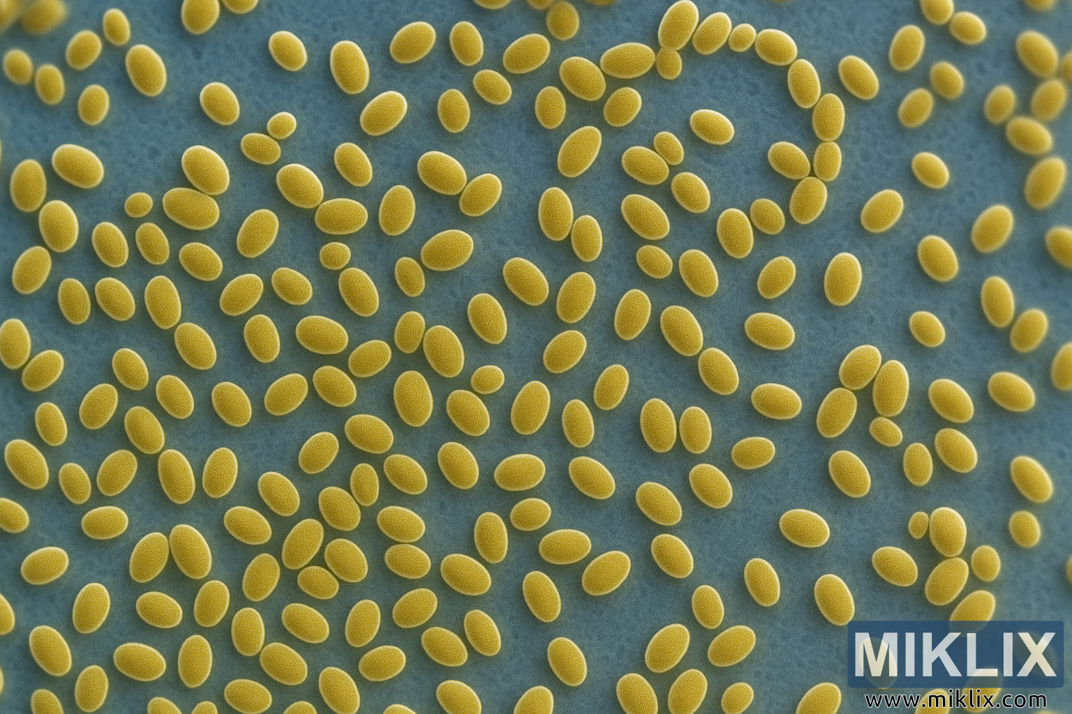
A detailed microscopic image of Saccharomyces cerevisiae var. diastaticus, highlighting its distinctive oval yeast cells with textured membranes and budding patterns under high magnification.

Available versions of this image
The image files available for download below are less compressed and higher resolution - and as a result of that, higher quality - than the images embedded in articles and pages on this website, which are more optimized for file size in order to reduce bandwidth consumption.
Regular size (1,536 x 1,024)
Large size (3,072 x 2,048)
Very large size (4,608 x 3,072)
Extra large size (6,144 x 4,096)
Comically large size (1,048,576 x 699,051)
- Still uploading... ;-)
Image description
This high-resolution microscopic image presents a magnified view of Saccharomyces cerevisiae var. diastaticus, a variant of brewer’s yeast known for its diastatic activity — the ability to ferment complex sugars that typical yeast strains cannot. The composition reveals hundreds of yeast cells scattered across the frame, each appearing as golden-yellow ovoid structures against a smooth, bluish background. The contrast between the warm tones of the cells and the cool background emphasizes both their structure and density, evoking the clarity of an electron or fluorescence micrograph.
Each individual yeast cell displays a distinct morphology characteristic of Saccharomyces cerevisiae: thick outer membranes with finely granular textures suggesting a roughened surface typical of yeast cell walls rich in polysaccharides and glycoproteins. The interior of several cells shows subtle variations in shading, reflecting intracellular organelles or cytoplasmic density gradients. Some cells appear in pairs or small clusters, hinting at recent or ongoing budding — the asexual reproductive process through which yeast multiply. These budding forms are most apparent where one cell appears slightly attached or elongated toward another, capturing a dynamic biological moment frozen under high magnification.
The image’s color palette, dominated by golden tones on a muted cyan background, is both visually striking and scientifically informative. It conveys a sense of depth and texture, reminiscent of modern colorized microscopy techniques that transform grayscale electron micrographs into visually interpretable educational images. The apparent three-dimensionality of the cells suggests a shallow depth of field, providing a sense of their volume and curvature as light falls unevenly across the cell surfaces.
Saccharomyces cerevisiae var. diastaticus is of particular interest in brewing and fermentation industries, where it can cause secondary fermentation or over-attenuation due to its ability to secrete glucoamylase, an enzyme that breaks down dextrins into fermentable sugars. This unique capability differentiates it from standard brewing yeasts and makes its identification under the microscope crucial in quality control and microbiological diagnostics. The image captures not only the aesthetic beauty of the microorganism but also serves as a visual reference for scientists studying its physiological and morphological characteristics.
In terms of artistic and compositional quality, the image balances scientific realism with aesthetic precision. The spatial distribution of the cells, some isolated and others forming gentle clusters or linear chains, conveys the natural randomness of yeast suspension cultures. The crispness of the cell outlines and the soft diffusion of background tones create a sense of microscopic depth that bridges the line between documentary science and visual art. Every detail, from the delicate grain of the cell surface to the faint shadows hinting at the microscopic plane, contributes to a lifelike portrayal of one of the most extensively studied eukaryotic microorganisms in modern biology.
The image is related to: Fermenting Beer with CellarScience Saison Yeast