
Billede: Laboratory Analysis of Swiss Chard Compounds and Health Benefits
Udgivet:
Sidst opdateret: 12. september 2026 kl. 14.49.56 UTC
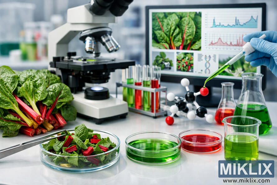
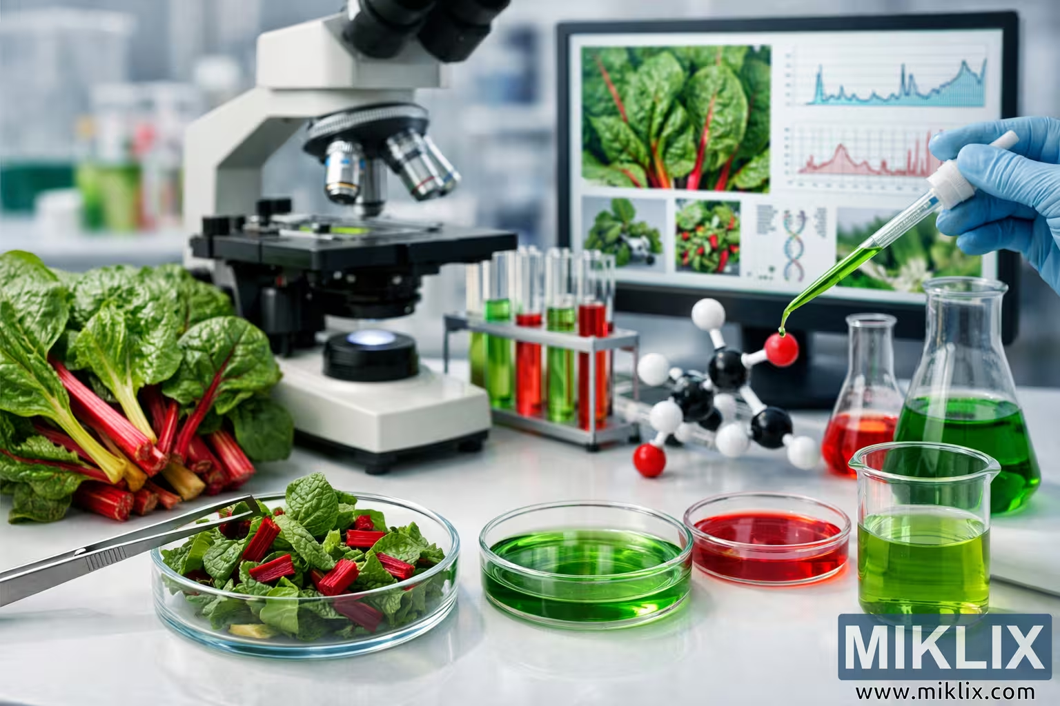
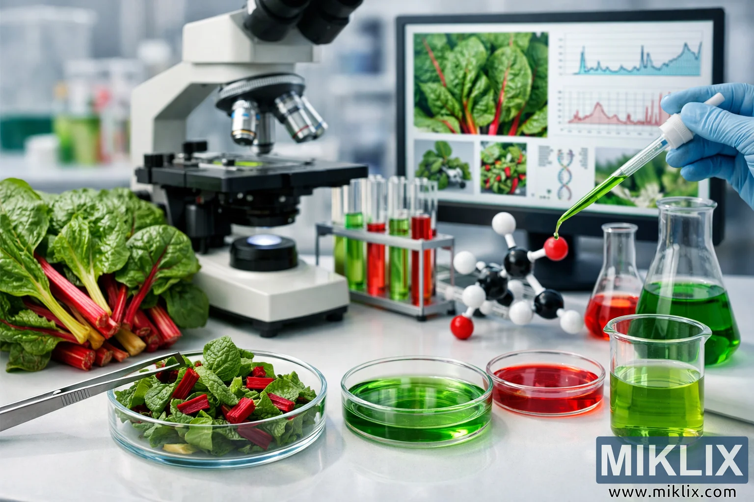
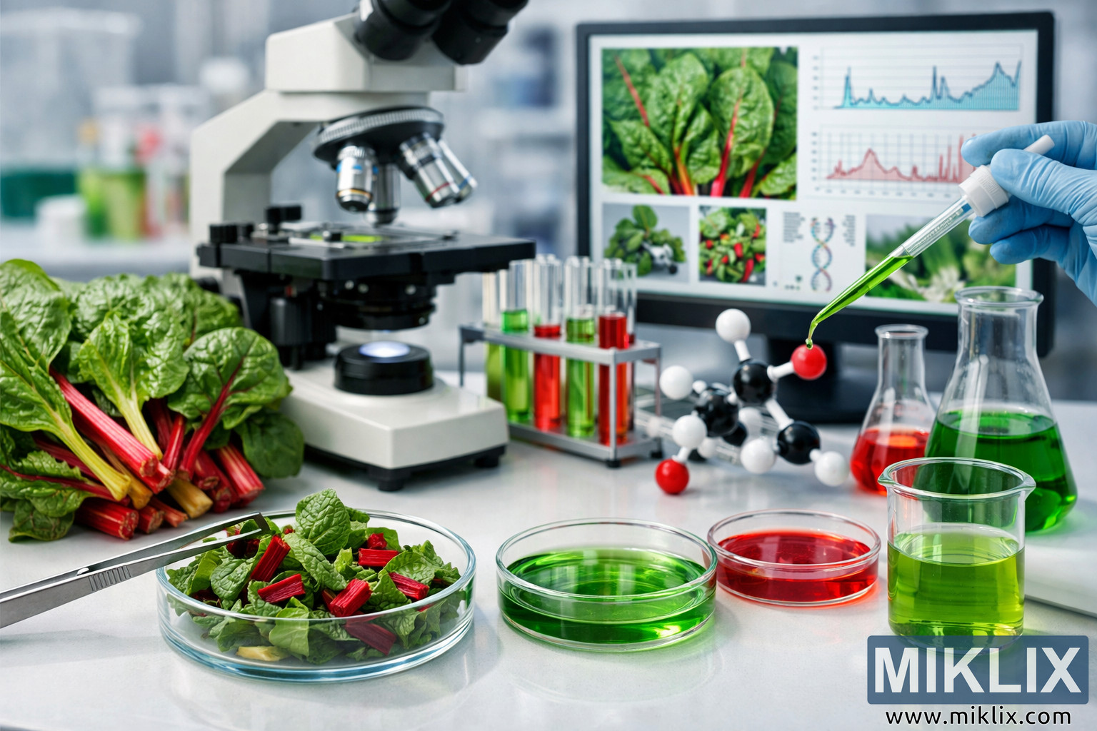
A detailed laboratory research scene featuring Swiss chard leaves, colorful plant extracts, test tubes, flasks, a microscope, molecular models, and a computer monitor displaying analytical data. The image highlights scientific investigation into Swiss chard compounds and their potential health benefits in a clean, modern lab environment.
Laboratory Analysis of Swiss Chard Compounds and Health Benefits
Tilgængelige versioner af dette billede
De billedfiler, der kan downloades nedenfor, er mindre komprimerede og har højere opløsning - og dermed højere kvalitet - end de billeder, der er indlejret i artikler og sider på dette websted, som er mere optimerede med hensyn til filstørrelse for at reducere båndbreddeforbruget.
Almindelig størrelse (1,536 x 1,024)
Stor størrelse (3,072 x 2,048)
Meget stor størrelse (4,608 x 3,072)
Ekstra stor størrelse (6,144 x 4,096)
Komisk stor størrelse (1,048,576 x 699,051)
- Uploader stadig... ;-)
Billedbeskrivelse
This image depicts a modern scientific research laboratory dedicated to studying the bioactive compounds and potential health benefits of Swiss chard. The scene is arranged on a wide, clean laboratory bench, with a careful balance between fresh plant material, analytical instruments, and colorful chemical solutions that visually communicate the intersection of nutrition science and laboratory research.
In the foreground on the left side, a clear glass Petri dish rests on the smooth white surface of the bench. Inside the dish, freshly cut Swiss chard leaves and stems are neatly arranged. The leaves are a rich, vibrant green, with visible veins and slightly crinkled textures that suggest freshness and high moisture content. The stems, sliced into short segments, display bright red hues, adding a striking contrast to the green foliage. Hovering just above the Petri dish, a pair of stainless steel tweezers holds a small piece of Swiss chard leaf, as if a scientist has just lifted it for closer inspection or is about to transfer it to another container for analysis. The metallic sheen of the tweezers and their precise grip emphasize the careful, methodical nature of the research.
To the right of this first Petri dish, two additional dishes are placed in a subtle diagonal line. One contains a vivid green liquid sample, smooth and translucent, suggesting an extract derived from the leafy portions of the Swiss chard. The other holds a deep red liquid, likely representing an extract rich in pigments or specific phytonutrients associated with the red stems. The liquids are evenly distributed across the surfaces of the dishes, reflecting the overhead lighting and hinting at the controlled conditions under which the samples are prepared. Together, these Petri dishes form a visual narrative of plant material being transformed into analyzable solutions.
On the far left side of the bench, a fresh bunch of Swiss chard lies partially overlapping the workspace. The leaves are large and lush, with crisp edges and a slightly glossy surface that catches the light. The stems range in color from bright red to warm yellow, creating a rainbow-like effect that underscores the plant’s natural diversity of pigments. This bunch of Swiss chard appears unprocessed, serving as a reference point for the raw material from which the laboratory samples are derived. Its presence reinforces the connection between the living plant and the scientific investigation taking place.
In the midground, a rack of test tubes stands upright, each tube containing liquids in varying shades of green and red. The test tubes are arranged in orderly rows, suggesting a systematic series of experiments or measurements. Some tubes hold pale, almost translucent green solutions, while others contain more saturated, darker tones. The red liquids range from bright crimson to deeper, wine-like hues. This gradient of colors implies different concentrations, extraction methods, or compound profiles being tested. The test tube rack itself is simple and functional, emphasizing the practical, methodical nature of the research environment.
To the right of the test tubes, several pieces of classic laboratory glassware are visible, including Erlenmeyer flasks and a graduated cylinder. One Erlenmeyer flask contains a green solution, while another holds a red solution, echoing the color scheme seen in the Petri dishes and test tubes. The graduated cylinder is filled with a clear liquid, possibly a solvent or distilled water used in the preparation of samples. The markings on the cylinder are faintly visible, indicating precise measurement capabilities. These vessels contribute to the sense of an active, ongoing experimental process, where liquids are mixed, measured, and analyzed to uncover the chemical composition of Swiss chard.
At the center of the image, a compound microscope stands prominently on the bench, its objective lenses pointed downward toward the stage. The microscope’s body is sturdy and professional, with multiple lenses suggesting different magnification levels. This instrument symbolizes the microscopic examination of Swiss chard tissues, cells, and structures, as well as the potential observation of extracted compounds at a cellular or subcellular level. The positioning of the microscope in the middle of the scene highlights its importance as a primary tool for detailed analysis.
In the foreground on the right side, a glass beaker holds a green liquid, similar in tone to the solutions seen elsewhere in the image. Next to the beaker, a molecular model is arranged on the bench. The model consists of black, white, and red spheres connected by gray rods, representing atoms and bonds in a simplified chemical structure. This model suggests that the research extends beyond simple observation, delving into the molecular architecture of Swiss chard compounds—perhaps antioxidants, vitamins, or other bioactive molecules that contribute to health benefits. The presence of the molecular model bridges the gap between the tangible plant material and the abstract chemical structures that underpin its nutritional value.
On the right side of the image, a computer monitor displays detailed data related to Swiss chard analysis. The screen shows high-resolution images of Swiss chard leaves under magnification, revealing intricate patterns of veins and cellular structures. Alongside these images, graphs and charts present chemical analysis results, such as concentration curves, bar charts, or line graphs that track the presence of specific compounds. A stylized DNA helix graphic appears on the screen, hinting at genetic or genomic aspects of the research, or at least the biological context in which these compounds operate. The monitor’s display reinforces the idea that the laboratory work integrates visual microscopy, quantitative data analysis, and biological interpretation.
In the midground near the monitor, additional laboratory tools are visible, including pipettes and a small amber dropper bottle. The pipettes suggest precise transfer of liquids between containers, while the amber bottle hints at light-sensitive reagents or extracts that must be protected from direct illumination. These items contribute to the authenticity of the laboratory setting, indicating that multiple techniques and instruments are employed in the research process.
On the far right, a scientist’s gloved hand is captured in the act of dispensing a green liquid into a test tube using a pipette. The glove is a light, neutral color, emphasizing hygiene and safety. The pipette is held steadily, and a small droplet of green solution is either entering or about to enter the test tube, symbolizing the careful, incremental steps that define laboratory experimentation. This human element adds a dynamic touch to the otherwise static arrangement of objects, reminding the viewer that behind every measurement and sample is a researcher actively engaged in discovery.
In the background, beyond the main bench and equipment, blurred laboratory instruments and containers can be seen, suggesting a larger research facility with multiple workstations and ongoing projects. The overall lighting in the laboratory is cool, bright, and evenly distributed, minimizing shadows and highlighting the clarity of the glassware, liquids, and plant material. The environment appears clean, organized, and professional, reinforcing the credibility of the scientific work being conducted.
The color palette of the image is dominated by the natural greens and reds of Swiss chard and its extracts, contrasted against the neutral whites and grays of the laboratory surfaces and equipment. This combination creates a visually engaging scene that communicates both the vibrancy of plant-based nutrition and the precision of scientific inquiry. The image as a whole tells a story of how Swiss chard, a humble leafy vegetable, becomes the subject of sophisticated research aimed at understanding its compounds, their molecular structures, and their potential contributions to human health.
Billedet er relateret til: Swiss Chard: Why It Is One of the Healthiest Leafy Greens