
Pilt: Anti-Inflammatory Pathway Illustration in the Human Body
Avaldatud:
Viimati uuendatud: 16. juuli 2026, kell 20:43:50 UTC
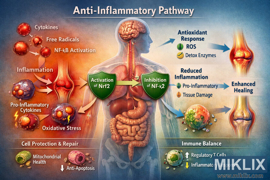
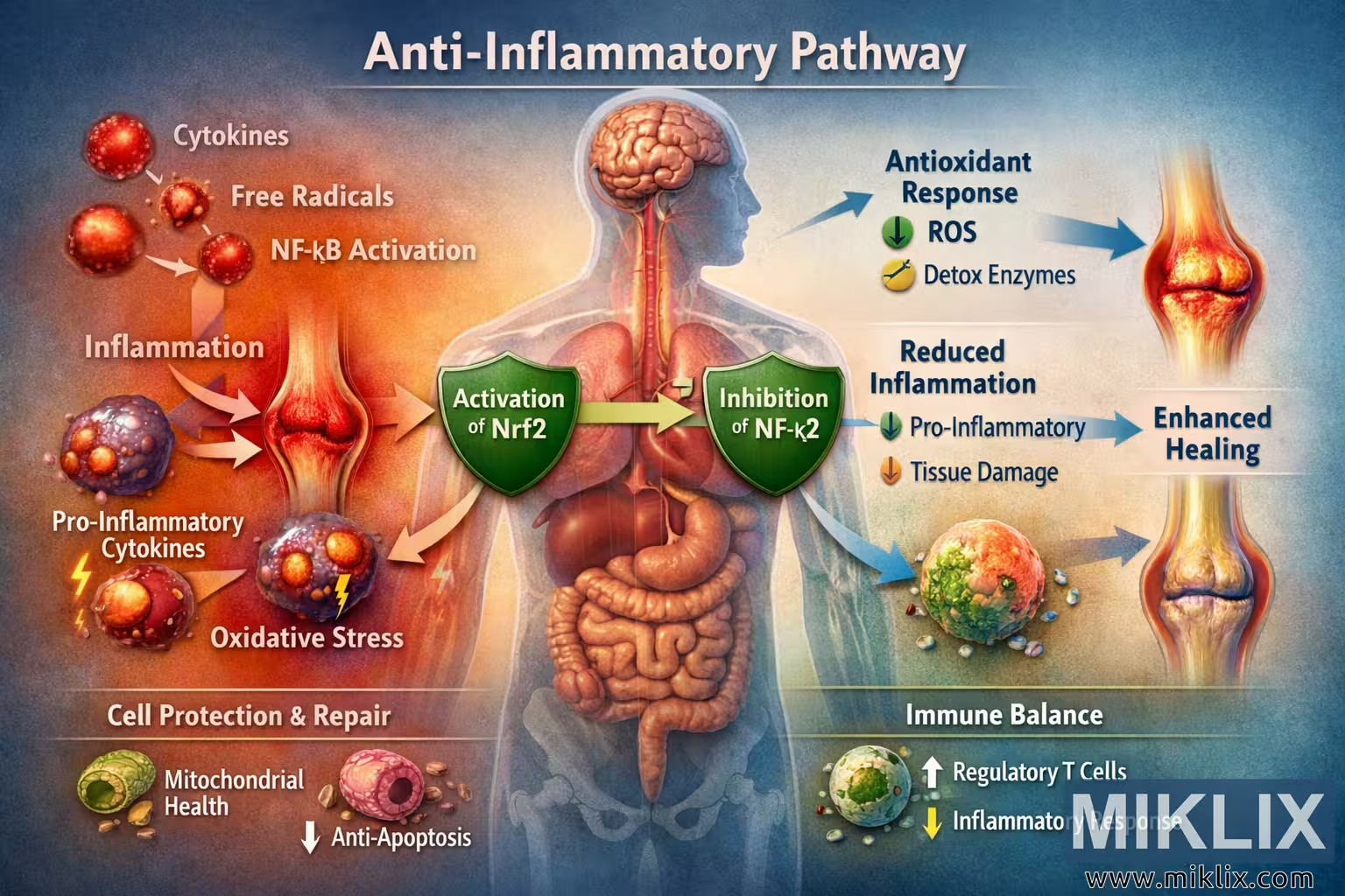
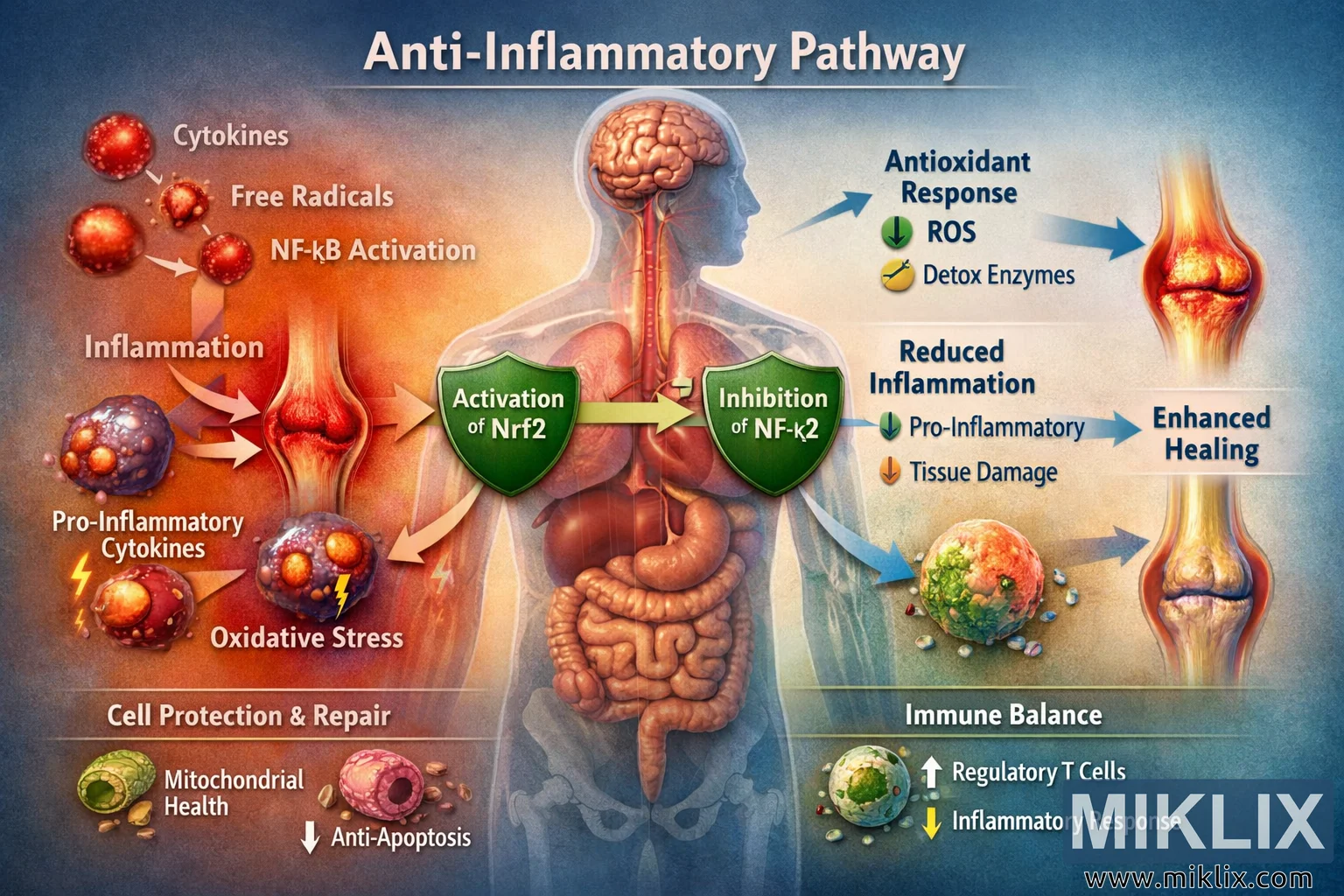
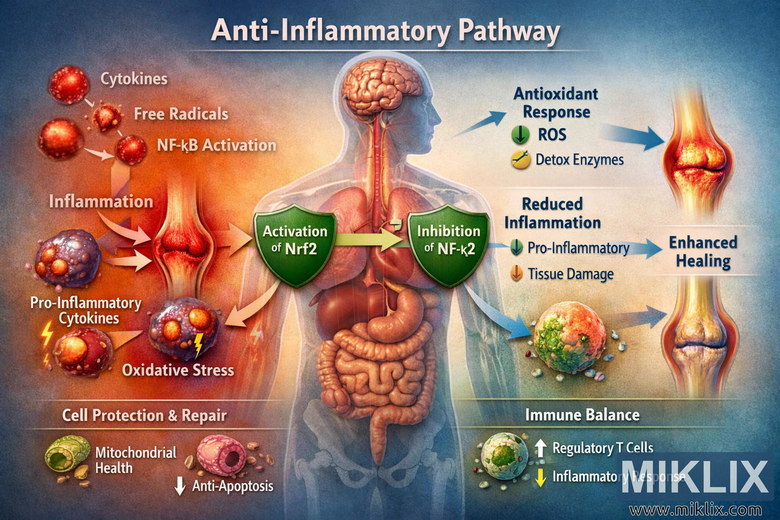
High-resolution educational illustration of the anti-inflammatory pathway in the human body, featuring a semi-transparent human silhouette, inflammatory factors, natural molecules, and key signaling steps such as Nrf2 activation and NF-κB inhibition. The diagram visually explains how antioxidant responses and reduced inflammatory cytokines lead to enhanced healing, cell protection, and immune balance.
Anti-Inflammatory Pathway Illustration in the Human Body
Selle pildi olemasolevad versioonid
Allpool allalaadimiseks saadaval olevad pildifailid on vähem kokkusurutud ja kõrgema resolutsiooniga - ja selle tulemusena kvaliteetsemad - kui selle veebisaidi artiklitesse ja lehekülgedele manustatud pildid, mis on ribalaiuse tarbimise vähendamiseks faili suuruse poolest rohkem optimeeritud.
Tavaline suurus (1,536 x 1,024)
Suur suurus (3,072 x 2,048)
Väga suur suurus (4,608 x 3,072)
Eriti suur suurus (6,144 x 4,096)
Koomiliselt suur suurus (1,048,576 x 699,051)
- Ikka veel üleslaadimine... ;-)
Pildi kirjeldus
This educational digital illustration presents a detailed visualization of the anti-inflammatory pathway within the human body, designed to help viewers understand how molecular interactions and cellular responses work together to reduce inflammation and promote healing. The composition is laid out in a clear, landscape orientation, with a semi-transparent human silhouette occupying the central portion of the image. Through this translucent figure, key internal organs are visible, including the brain, lungs, heart, and intestines, symbolizing the systemic nature of inflammation and anti-inflammatory responses throughout the body.
At the very top of the illustration, the title "Anti-Inflammatory Pathway" appears in bold, white, capitalized letters, standing out against a darker, gradient background. This heading immediately frames the image as a scientific and educational diagram. The color palette of the background transitions smoothly from deep blue and teal tones on the right—representing calm, healing, and anti-inflammatory effects—to warmer red and orange hues on the left—representing inflammation, stress, and cellular damage. This gradient subtly reinforces the conceptual journey from inflammation to resolution.
On the left side of the image, several red circular icons represent key inflammatory factors. Each icon is labeled with white text: "Cytokines," "Free Radicals," and "NF-κB Activation." These icons are arranged vertically, with thin, bright arrows pointing inward toward the center of the illustration. The red color of these icons conveys danger and cellular stress, while their circular shape suggests discrete molecular entities or signaling hubs. Near these icons, additional visual cues such as jagged lightning bolts and small flame-like shapes emphasize the destructive potential of uncontrolled inflammation.
Following the arrows from the left, the viewer’s eye is drawn to a central depiction of an inflamed joint, shown with pronounced redness, swelling, and a glowing halo of irritation around the joint area. This joint is labeled "Inflammation" in clear text. The joint serves as a focal example of how systemic inflammatory signals can manifest as localized pain and tissue damage. Just below this joint, two stylized cells are depicted. One is labeled "Pro-Inflammatory Cytokines" and the other "Oxidative Stress." From these cells, red lightning bolts and fiery icons emanate outward, visually representing the release of harmful mediators that exacerbate inflammation and damage surrounding tissues.
In the central region of the illustration, the narrative shifts from harmful factors to protective, anti-inflammatory agents. Here, three icons representing natural molecules are displayed: "Omega-3," "Curcumin," and "Polyphenols." Each of these is depicted as a colored capsule or droplet, with soft gradients of blue, green, and golden yellow. These molecules are arranged in a cluster, with arrows leading from them toward two prominent green shield icons. The shields are labeled "Activation of Nrf2" and "Inhibition of NF-κB." The shields symbolize defense and protection, visually communicating that these pathways help guard cells against oxidative damage and excessive inflammatory signaling.
The "Activation of Nrf2" shield is associated with a series of downstream effects. From this shield, a bright green arrow extends to a section labeled "Antioxidant Response." In this section, smaller icons and labels appear, including "↓ ROS" (indicating reduced reactive oxygen species) and "Detox Enzymes." These icons are colored in cool greens and blues, with subtle sparkles and clean, rounded shapes that suggest cleansing and restoration. The overall effect is to show that activating Nrf2 leads to enhanced antioxidant defenses, helping cells neutralize free radicals and reduce oxidative stress.
Similarly, the "Inhibition of NF-κB" shield connects via a green arrow to a section labeled "Reduced Inflammation." Here, icons and labels such as "↓ Pro-Inflammatory Cytokines" and "↓ Tissue Damage" are displayed. The icons in this area are calmer and more subdued compared to the fiery shapes on the left side of the image. They use softer colors and smoother lines, visually indicating that inflammatory signals are being dampened and tissue injury is being minimized. Together, these two shield pathways—Nrf2 activation and NF-κB inhibition—form the central conceptual bridge from harmful stimuli to protective responses.
To the far right of the illustration, the pathway culminates in a section labeled "Enhanced Healing." Arrows from both the antioxidant and anti-inflammatory sections converge here, symbolizing that these processes work together to restore tissue health. Within this area, icons and labels such as "↓ Pain," "↓ Swelling," and "Tissue Repair" are shown. The visual representation of a healed joint appears here, contrasting with the inflamed joint on the left. The healed joint is depicted with normal coloration, smooth contours, and a subtle glow of wellness, emphasizing the successful resolution of inflammation.
At the bottom of the image, two additional sections provide further detail on the downstream benefits of the anti-inflammatory pathway. On the left, a section labeled "Cell Protection & Repair" includes icons and text such as "Mitochondrial Health" and "Anti-Apoptosis." A stylized green mitochondrion is depicted, with gentle waves of energy surrounding it, symbolizing improved cellular energy production and resilience. The "Anti-Apoptosis" label suggests that cells are being protected from premature death, supporting tissue integrity and function.
On the right side of the bottom portion, a section labeled "Immune Balance" highlights the role of regulatory immune cells in maintaining homeostasis. Icons and labels such as "↑ Regulatory T Cells" and "↓ Inflammatory Response" are shown, along with a stylized T cell rendered in green and blue tones. This section visually communicates that the anti-inflammatory pathway does not simply suppress immunity, but rather fine-tunes it, promoting a balanced response that prevents chronic inflammation while preserving necessary defense mechanisms.
Throughout the illustration, arrows and labels connect each step of the pathway in a logical sequence. The flow begins with inflammatory triggers like cytokines, free radicals, and NF-κB activation, moves through the introduction of natural molecules such as omega-3, curcumin, and polyphenols, and then proceeds to the activation of Nrf2 and inhibition of NF-κB. From there, the diagram shows the resulting antioxidant response, reduced inflammation, and enhanced healing, culminating in improved cell protection, repair, and immune balance. The careful use of color—red for inflammation and damage, green and blue for protection and healing—helps viewers intuitively grasp the transition from a harmful state to a restored, healthy condition.
The human silhouette in the center serves as a unifying backdrop, reminding viewers that these molecular and cellular events occur within the context of the whole body. The visible organs suggest that inflammation and anti-inflammatory processes can affect multiple systems, including the nervous, respiratory, cardiovascular, and digestive systems. The overall design is clean, modern, and visually engaging, making the complex biology of anti-inflammatory pathways more accessible to students, healthcare professionals, and anyone interested in understanding how the body defends itself against chronic inflammation.
The image is rendered with high resolution and crisp detail, ensuring that all text labels, icons, and arrows are sharp and legible. The lighting is even and well-balanced, with no harsh shadows or overly bright highlights, allowing viewers to focus on the conceptual relationships rather than on dramatic visual effects. The depth of field is effectively infinite, keeping every element—from the smallest icon to the largest organ—clear and in focus. This clarity supports the educational purpose of the illustration, making it suitable for use in presentations, textbooks, websites, and patient education materials.
In summary, this anti-inflammatory pathway illustration in the human body provides a comprehensive, visually structured overview of how natural molecules can modulate key signaling pathways, reduce oxidative stress and inflammatory cytokines, and ultimately promote healing, cell protection, and immune balance. By combining a semi-transparent human figure with labeled icons, arrows, and color-coded sections, the image transforms complex biochemical processes into an intuitive visual narrative that guides the viewer from inflammation to resolution.
Pilt on seotud: Health Benefits of Chives: Nature's Nutritional Powerhouse