
تصویر: Corn Leaf with Gray Leaf Spot Disease Lesions
منتشر شده:
آخرین به روز رسانی: ۱۳ مارس ۲۰۲۶ ساعت ۲۰:۳۸:۴۴ (UTC)
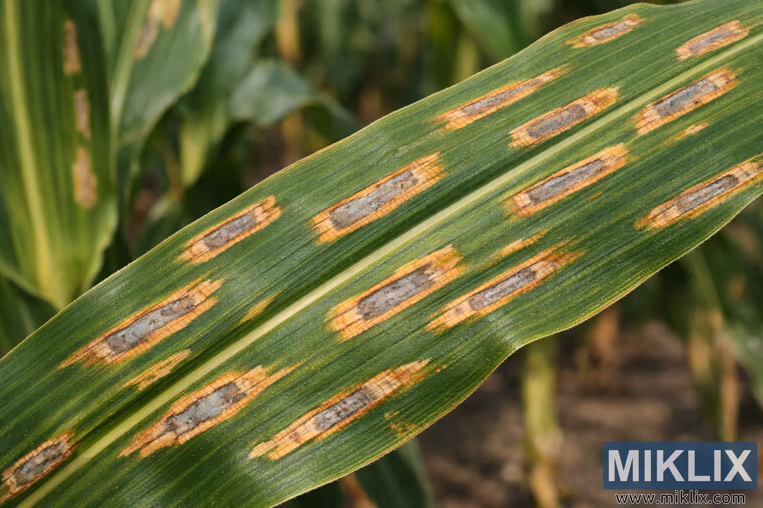
High-resolution close-up of a corn leaf affected by gray leaf spot disease, showing characteristic rectangular lesions aligned with the leaf veins.
Corn Leaf with Gray Leaf Spot Disease Lesions

نسخههای موجود از این تصویر
فایلهای تصویری که در زیر برای دانلود موجود است، نسبت به تصاویر موجود در مقالات و صفحات این وبسایت که برای کاهش مصرف پهنای باند، از نظر حجم فایل بهینهتر شدهاند، فشردهسازی کمتری داشته و وضوح بالاتری دارند - و در نتیجه، کیفیت بالاتری دارند.
اندازه معمولی (1,536 x 1,024)
سایز بزرگ (3,072 x 2,048)
سایز بسیار بزرگ (4,608 x 3,072)
سایز فوق العاده بزرگ (6,144 x 4,096)
اندازهی کمیک بزرگ (1,048,576 x 699,051)
- هنوز در حال آپلود هستم... ;-)
شرح تصویر
This high-resolution landscape photograph presents a detailed close-up view of a corn (maize) leaf exhibiting classic symptoms of gray leaf spot disease. The leaf stretches diagonally across the frame from the lower left toward the upper right, dominating the composition and drawing the viewer’s attention to the distinctive lesions scattered across its surface. The leaf is deep green with clearly visible parallel veins that run lengthwise, forming the structural pattern typical of maize foliage. Along these veins appear multiple elongated, rectangular lesions that are characteristic of gray leaf spot disease, a fungal infection caused by Cercospora species.
Each lesion is aligned parallel to the leaf veins and varies slightly in size, but most are elongated rectangles with fairly straight sides and rounded ends. The centers of the lesions appear gray to light tan, giving them a somewhat bleached or ashen appearance compared with the surrounding healthy tissue. Around many of the lesions, the leaf tissue shows a subtle yellow to pale brown halo that transitions into the healthy green areas of the leaf. The borders between infected and healthy tissue are fairly distinct, highlighting the geometric shape of the disease spots.
The lesions are distributed irregularly across the leaf blade but tend to follow the orientation of the veins, creating a repeating pattern of rectangular marks that emphasize the linear structure of the leaf. Some lesions appear isolated, while others are clustered closer together, suggesting areas where the disease has progressed more extensively. The surface texture of the leaf is clearly visible, with fine ridges and a slightly glossy appearance where sunlight reflects off the healthy tissue.
The background is softly blurred, creating a shallow depth-of-field effect that keeps the diseased leaf sharply in focus. Out-of-focus corn plants, stalks, and hints of soil form a natural agricultural setting, suggesting that the photograph was taken in a corn field during the growing season. The blurred green shapes of neighboring leaves and the muted brown tones of the soil provide contextual information without distracting from the primary subject.
Lighting appears natural and even, likely captured outdoors under daylight conditions. The illumination highlights the contrast between the green leaf tissue and the grayish lesions, making the disease symptoms easy to observe and study. The combination of sharp focus, natural color, and high resolution allows viewers to clearly examine the disease characteristics, making the image suitable for educational, diagnostic, or agricultural reference purposes.
تصویر مربوط به: Growing Corn: Your Complete Guide to Sweet Success in the Garden