
תְמוּנָה: Plant Cell Cultures in Laboratory Petri Dishes
פורסם:
עודכן לאחרונה: 22 במאי 2026 בשעה 18:03:45 UTC
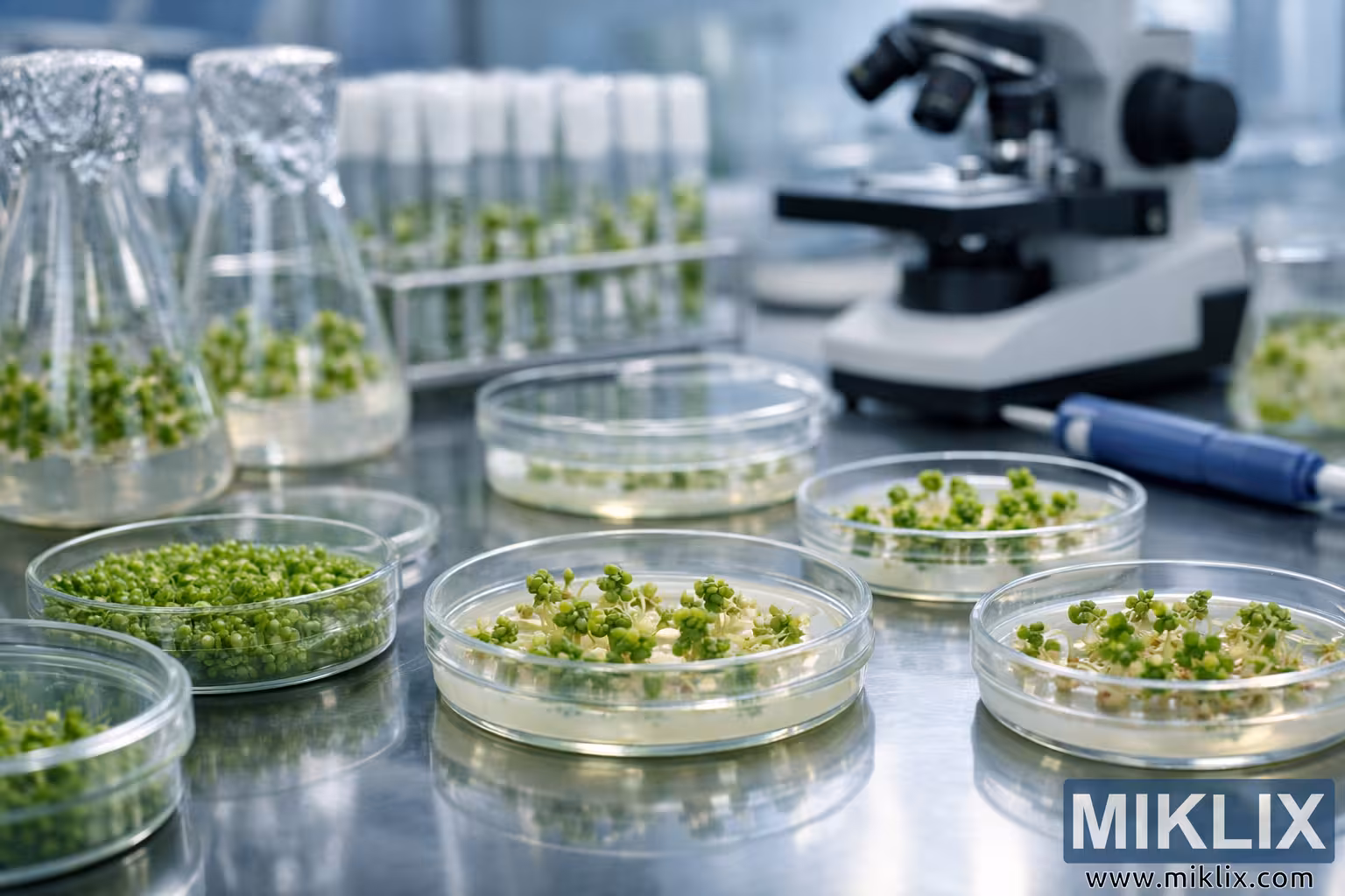
A detailed laboratory scene showing Petri dishes with plant cell cultures, flasks, and scientific equipment under bright lighting—illustrating modern plant biotechnology research.
Plant Cell Cultures in Laboratory Petri Dishes

גרסאות זמינות של תמונה זו
קבצי התמונה הזמינים להורדה להלן דחוסים פחות ובעלי רזולוציה גבוהה יותר - וכתוצאה מכך, באיכות גבוהה יותר - בהשוואה לתמונות המוטמעות במאמרים ובדפים באתר זה, אשר מותאמות יותר לגודל הקובץ על מנת להפחית את צריכת רוחב הפס.
גודל רגיל (1,536 x 1,024)
גודל גדול (3,072 x 2,048)
גודל גדול מאוד (4,608 x 3,072)
גודל גדול במיוחד (6,144 x 4,096)
גודל גדול באופן קומי (1,048,576 x 699,051)
- עדיין מעלה... ;-)
תיאור התמונה
This image captures a meticulously arranged laboratory environment dedicated to plant biotechnology and tissue culture research. The composition is dominated by a series of transparent Petri dishes placed on a reflective stainless steel surface, each containing vibrant green plant cell cultures in various stages of development. The foreground features several Petri dishes filled with agar-based nutrient media, where clusters of plant cells have begun to differentiate into small shoots and callus tissues. These tiny green structures, some with delicate leaves emerging, symbolize the controlled regeneration process that scientists use to study plant growth and genetic expression.
To the left, a group of Erlenmeyer flasks wrapped with breathable aluminum foil rests on the same surface. Inside each flask, small plantlets float in a clear liquid medium, their roots and stems visible through the glass. The foil coverings indicate sterile conditions, essential for maintaining uncontaminated cultures. Behind the flasks, a rack of test tubes stands upright, each containing miniature plant samples suspended in transparent gel media. The test tubes are capped with white plugs, ensuring isolation from airborne particles while allowing limited gas exchange.
On the right side of the frame, a laboratory microscope is partially visible, its lenses and stage slightly blurred due to the shallow depth of field. This optical instrument represents the analytical side of the experiment—where scientists examine cellular morphology and growth patterns under magnification. A blue pipette with a digital adjustment display lies nearby, ready for precise measurement and transfer of liquids.
The lighting in the laboratory is bright, cool, and evenly distributed, emphasizing the sterile and controlled atmosphere typical of scientific research facilities. The reflective surface of the table creates subtle mirror effects, doubling the visual impact of the Petri dishes and flasks. The background fades into a soft blur, drawing attention to the intricate details of the plant cultures in the foreground.
Each Petri dish tells a different story of cellular transformation. One dish contains a dense carpet of green cells forming a uniform layer across the agar surface, while another shows scattered clusters of callus tissue beginning to organize into structured plantlets. The diversity of growth stages highlights the complexity of plant tissue culture—a process that involves inducing totipotent cells to regenerate entire plants under controlled conditions.
The overall aesthetic of the image is clean, modern, and scientific. The interplay of glass, metal, and organic material creates a balance between technology and nature. The cool tones of the laboratory environment contrast beautifully with the vivid greens of the plant cultures, symbolizing the harmony between human innovation and biological life.
This photograph not only documents a scientific procedure but also evokes a sense of wonder about the microscopic processes that sustain plant life. It represents the intersection of biology, chemistry, and environmental science, where researchers explore sustainable methods for crop improvement, genetic modification, and conservation. The image’s clarity, composition, and lighting make it an ideal visual representation of modern plant biotechnology and laboratory precision.
התמונה קשורה ל: A Guide to the Health Benefits of Dandelions