
Gambar: Corn Leaf with Stewart’s Bacterial Wilt Lesions
Diterbitkan:
Terakhir diperbarui: 13 Maret 2026 pukul 20.38.25 UTC
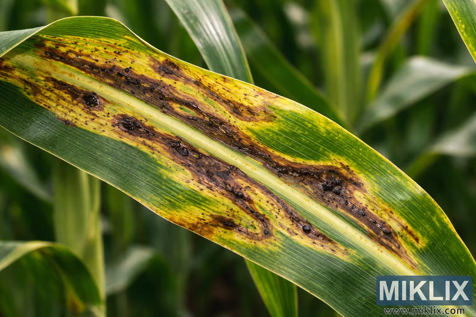
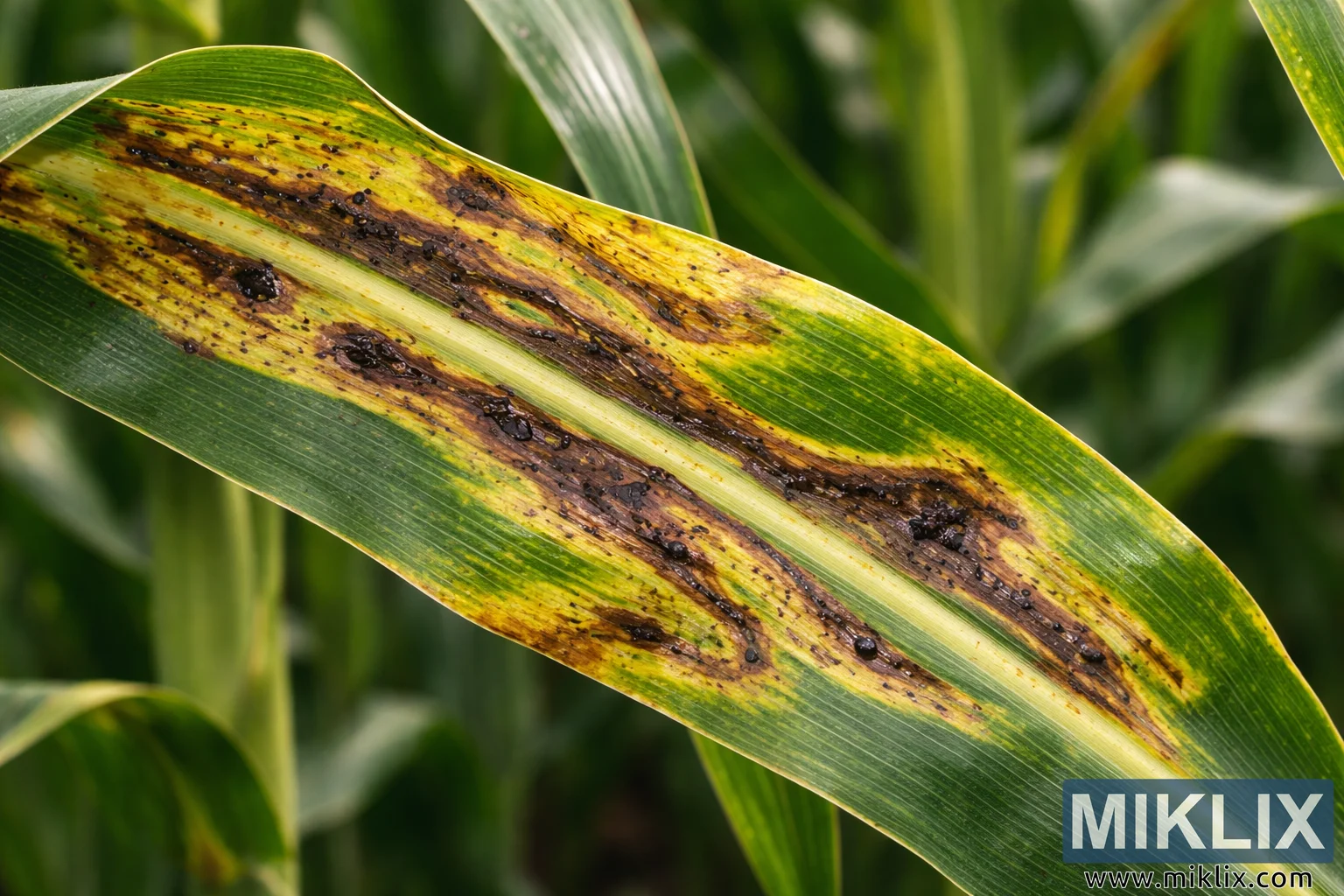
Detailed close-up image of a corn leaf affected by Stewart’s bacterial wilt, displaying elongated yellow-brown lesions with dark bacterial exudates and chlorotic halos.
Corn Leaf with Stewart’s Bacterial Wilt Lesions

Versi yang tersedia dari gambar ini
File gambar yang tersedia untuk diunduh di bawah ini tidak terlalu terkompresi dan beresolusi lebih tinggi - dan sebagai hasilnya, kualitasnya lebih tinggi - daripada gambar yang disematkan pada artikel dan halaman di situs web ini, yang lebih dioptimalkan untuk ukuran file guna mengurangi konsumsi bandwidth.
Ukuran biasa (1,536 x 1,024)
Ukuran besar (3,072 x 2,048)
Ukurannya sangat besar (4,608 x 3,072)
Ukuran ekstra besar (6,144 x 4,096)
Ukuran besar secara komik (1,048,576 x 699,051)
- Masih mengunggah... ;-)
Deskripsi gambar
This high-resolution close-up image shows a single corn (maize) leaf displaying clear and advanced symptoms of Stewart’s bacterial wilt, a disease caused by the bacterium Pantoea stewartii. The leaf is oriented diagonally across the frame from the lower left to the upper right, occupying most of the image. Its natural green color is interrupted by a series of elongated, irregular lesions that run parallel to the leaf veins. These lesions appear yellowish to tan at the edges and dark brown in the center, forming streak-like patterns typical of the disease’s progression along vascular tissue. Surrounding many of the lesions are distinct chlorotic halos—bright yellow areas where the plant tissue has begun to lose chlorophyll due to infection.
Within the darker central portions of several lesions, small black, glossy droplets and speckled accumulations are visible. These represent bacterial exudates and degraded plant material that collect as the pathogen multiplies and spreads through the leaf tissue. The lesions vary in size and shape; some remain narrow and elongated, while others have merged together into wider necrotic streaks. In certain areas the infection appears more advanced, where the tissue has turned a deeper brown and the leaf surface looks slightly sunken or deteriorated.
The leaf’s central midrib remains visible as a pale, raised line running through the middle, providing contrast with the diseased tissue surrounding it. Parallel veins characteristic of corn foliage create a linear texture that emphasizes how the infection follows vascular pathways. Despite the damage, some sections of the leaf still retain healthy green coloration, highlighting the stark difference between living tissue and areas affected by the bacterial disease.
The background is softly blurred, consisting of out-of-focus green corn leaves and stems. This shallow depth of field isolates the infected leaf as the main subject and provides context that the plant is located within a corn field. The lighting is natural and even, suggesting an outdoor agricultural setting. Subtle highlights on the leaf surface reveal its waxy texture and the slightly raised edges around some lesions.
Overall, the photograph provides a clear visual example of Stewart’s bacterial wilt symptoms on corn foliage. The combination of elongated yellow-bordered lesions, dark necrotic streaks, and visible bacterial exudates demonstrates the characteristic appearance of the disease as it spreads along the leaf veins. The image is well suited for educational, diagnostic, or agricultural reference purposes, helping viewers recognize the distinctive patterns associated with this bacterial infection in maize crops.
Gambar terkait dengan: Growing Corn: Your Complete Guide to Sweet Success in the Garden