
Gambar: Cancer Cells and Antioxidant Protection Visualization
Diterbitkan:
Terakhir diperbarui: 30 Juli 2026 pukul 18.37.23 UTC
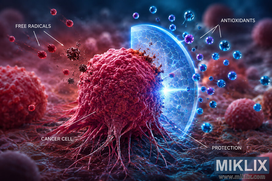
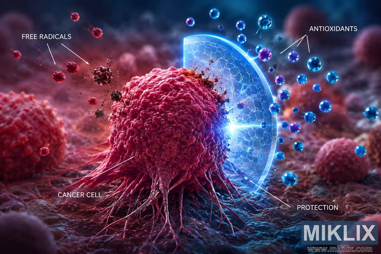
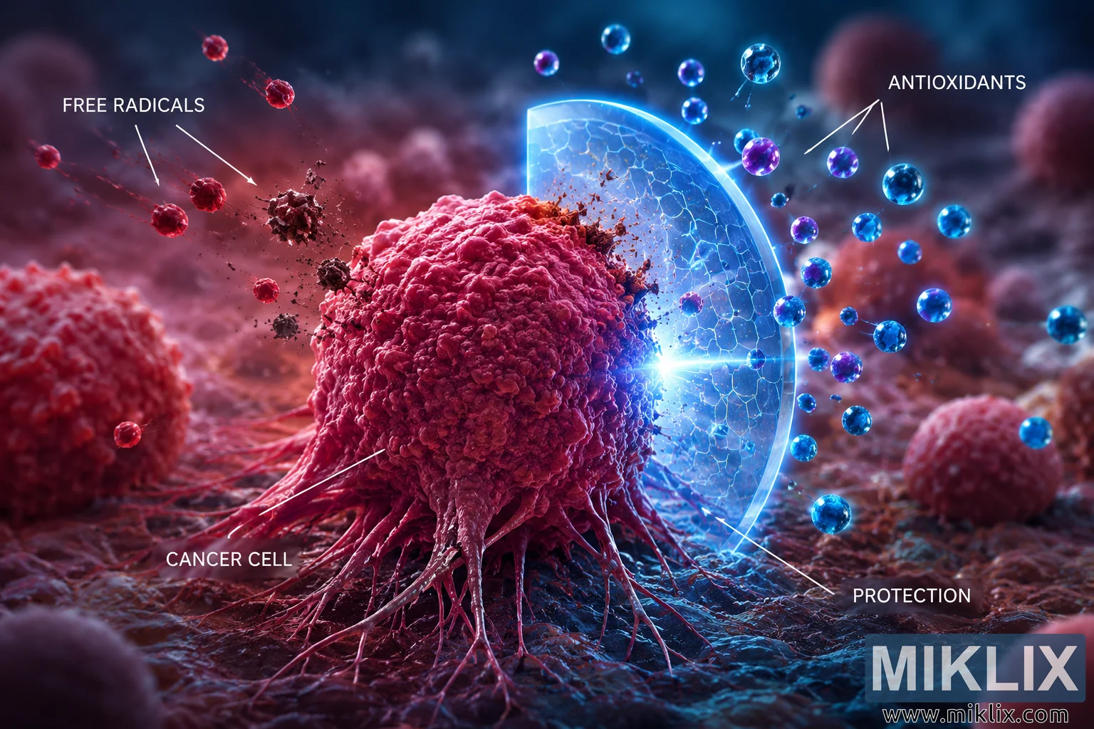
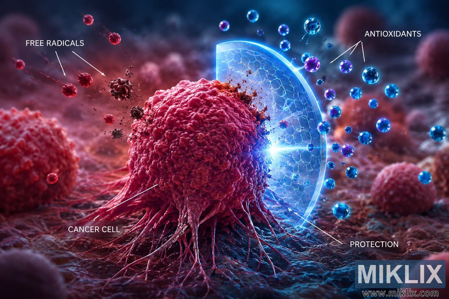
High-detail 3D medical illustration visualizing the interaction between free radicals, antioxidants, and a cancer cell. The conceptual artwork contrasts oxidative damage with antioxidant-mediated protection using glowing particles and a transparent defensive shield.
Cancer Cells and Antioxidant Protection Visualization
Versi yang tersedia dari gambar ini
File gambar yang tersedia untuk diunduh di bawah ini tidak terlalu terkompresi dan beresolusi lebih tinggi - dan sebagai hasilnya, kualitasnya lebih tinggi - daripada gambar yang disematkan pada artikel dan halaman di situs web ini, yang lebih dioptimalkan untuk ukuran file guna mengurangi konsumsi bandwidth.
Ukuran biasa (1,536 x 1,024)
Ukuran besar (3,072 x 2,048)
Ukurannya sangat besar (4,608 x 3,072)
Ukuran ekstra besar (6,144 x 4,096)
Ukuran besar secara komik (1,048,576 x 699,051)
- Masih mengunggah... ;-)
Deskripsi gambar
This landscape-oriented scientific illustration presents a highly detailed conceptual visualization of the relationship between cancer cells, free radicals, and antioxidant protection. The composition is designed as an educational medical graphic rather than a literal microscopic photograph, using realistic three-dimensional rendering techniques to communicate biological concepts through symbolic visual elements. The overall color palette contrasts warm reds and magentas with cool blues and cyans, emphasizing the opposing forces represented within the scene.
Dominating the center of the composition is a large, irregularly shaped cancer cell rendered with remarkable surface detail. The cell exhibits an uneven, highly textured membrane characterized by folds, ridges, nodules, and organic protrusions that create the impression of an aggressive and abnormal cellular structure. Numerous elongated extensions project downward from the base of the cell, resembling invasive cellular processes that anchor it to the surrounding tissue environment. The cell's coloration consists primarily of deep crimson, magenta, and pink hues with subtle variations in brightness that emphasize depth, curvature, and biological complexity. Carefully placed highlights and shadows reinforce the three-dimensional appearance of the membrane while suggesting a moist, living biological surface.
The surrounding environment resembles a stylized microscopic tissue landscape with a shallow depth of field. The foreground displays an uneven organic surface textured with subtle ridges, soft folds, and biological material that appears consistent with living tissue. Additional spherical cellular structures are intentionally blurred in both the foreground and background, reinforcing the impression that the viewer is observing one focal cell within a larger biological environment. The background transitions smoothly between dark reds, purples, and cool blue tones while remaining softly out of focus, directing visual attention toward the central subject.
The left portion of the image represents oxidative stress. Multiple small red particles travel diagonally toward the cancer cell from the upper left, suggesting movement and impact. These particles symbolize free radicals and are illustrated as glowing red spheres surrounded by faint motion trails that communicate dynamic movement. Near the point where several particles approach the cancer cell, darker fragmented debris appears suspended in space, visually implying cellular damage or molecular disruption. White annotation lines point toward these incoming particles, accompanied by the label identifying them as free radicals. The arrangement creates a clear directional flow from the upper left toward the central cancer cell, reinforcing the narrative of oxidative attack.
In sharp visual contrast, the right side of the illustration depicts a conceptual representation of antioxidant activity. Numerous luminous blue and violet spherical particles float around the right half of the image. These particles vary in size and intensity, creating depth while conveying an energetic, protective presence. Positioned directly between these particles and the cancer cell is a large translucent shield rendered with a subtle hexagonal pattern across its surface. The shield emits a cool blue glow along its edges and appears semi-transparent, allowing portions of the cancer cell and surrounding tissue to remain visible through its surface. Bright illumination radiates from the center of the shield where it faces the cancer cell, symbolizing defensive protection against oxidative influences.
Several annotation lines identify the antioxidant particles and the protective barrier. The typography is clean and modern, using white text that remains clearly legible against the darker background. The labels identify the conceptual elements without overwhelming the artwork, allowing the scientific illustration to function as an educational figure suitable for presentations, articles, or teaching materials.
Lighting plays a significant role in communicating the intended contrast. Warm illumination dominates the left side of the scene, complementing the red free radical particles and damaged appearance near the impact region. Cooler blue illumination defines the right side, reflecting from the protective shield and antioxidant particles while casting subtle blue highlights across the adjacent portion of the cancer cell. This complementary lighting scheme visually separates the opposing concepts of oxidative damage and antioxidant defense while maintaining overall compositional harmony.
The illustration employs cinematic depth of field, realistic volumetric lighting, soft atmospheric haze, and high-resolution texturing to create a polished scientific visualization. Every component is intentionally arranged to guide the viewer's attention from the attacking free radicals on the left, through the central cancer cell, toward the protective antioxidant shield on the right. The resulting composition serves as a conceptual educational graphic illustrating the balance between oxidative stress and antioxidant activity. While inspired by biological processes, the shield and particle effects are symbolic artistic representations intended to communicate scientific ideas visually rather than depict literal cellular mechanisms. The overall image combines medical illustration aesthetics with modern digital rendering techniques to produce a visually compelling educational visualization suitable for scientific communication, healthcare education, editorial publications, and biomedical presentations.
Gambar terkait dengan: Okra Benefits: How This Nutritious Vegetable Supports Your Health