
Gambar: Microscopic View of Antioxidant Compounds in Purple Radicchio Cells
Diterbitkan:
Terakhir diperbarui: 8 September 2026 pukul 08.40.12 UTC
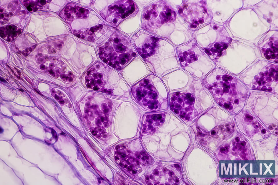
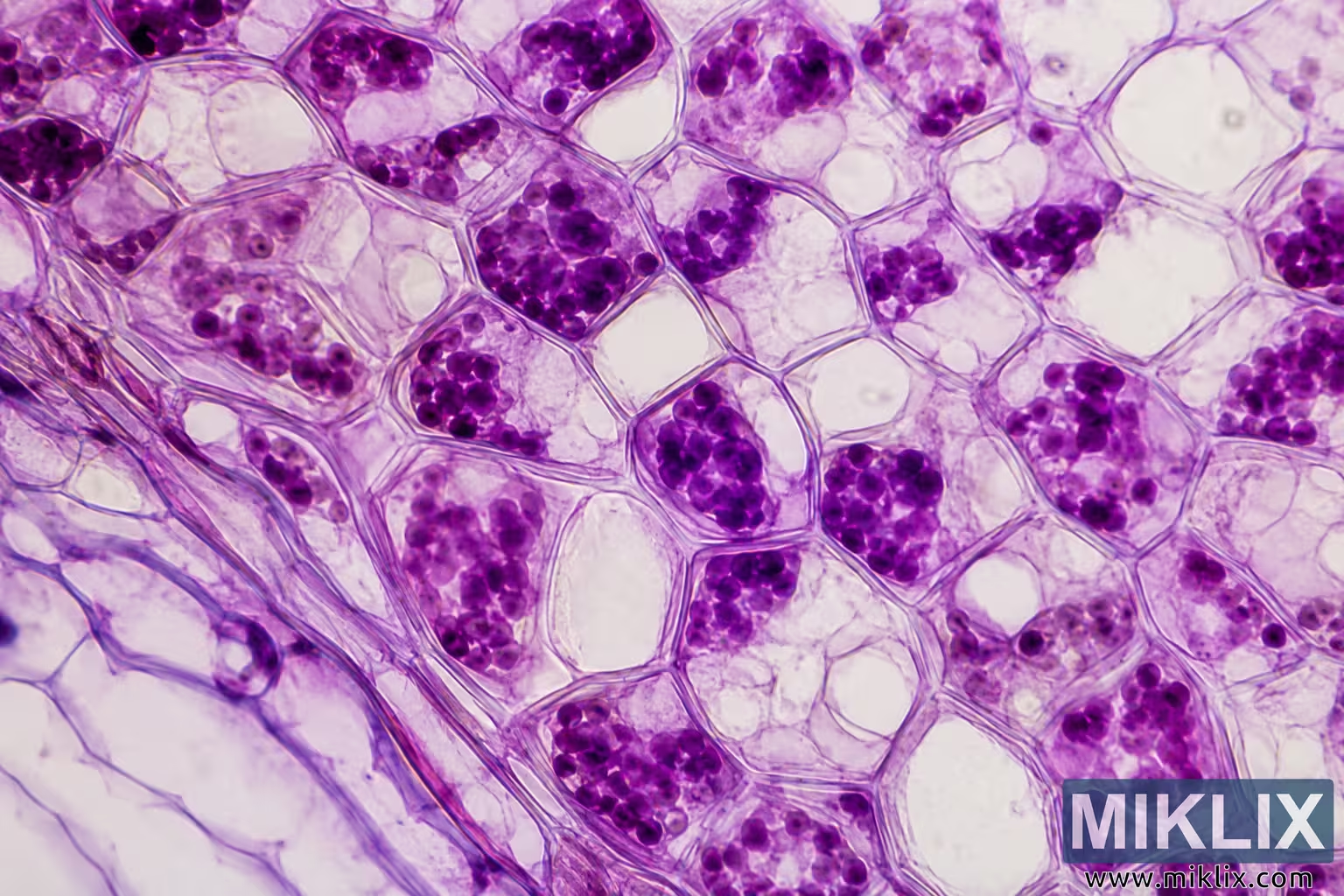
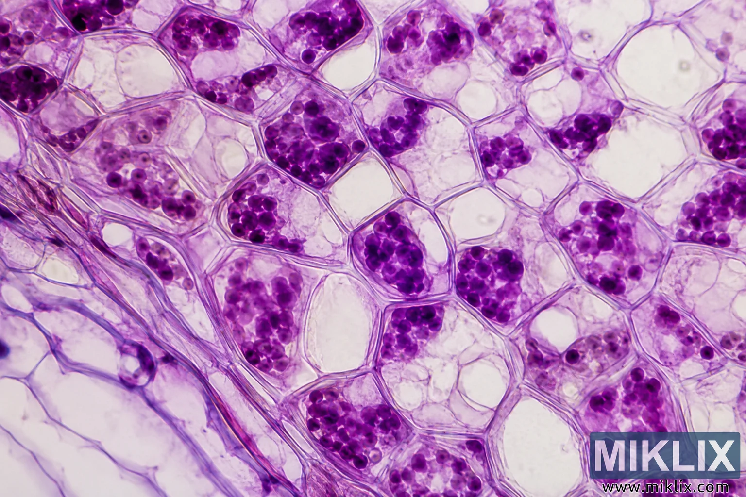
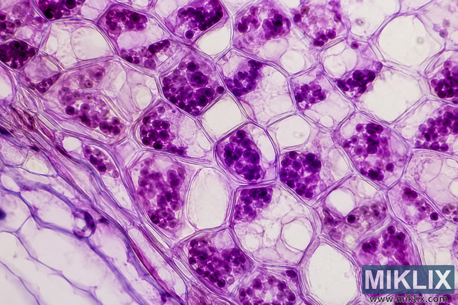
Detailed microscopic illustration of purple radicchio cells featuring vivid antioxidant-rich purple pigments, clearly defined plant cell walls, and intricate cellular structures in a high-resolution scientific style.
Microscopic View of Antioxidant Compounds in Purple Radicchio Cells
Versi yang tersedia dari gambar ini
File gambar yang tersedia untuk diunduh di bawah ini tidak terlalu terkompresi dan beresolusi lebih tinggi - dan sebagai hasilnya, kualitasnya lebih tinggi - daripada gambar yang disematkan pada artikel dan halaman di situs web ini, yang lebih dioptimalkan untuk ukuran file guna mengurangi konsumsi bandwidth.
Ukuran biasa (1,536 x 1,024)
Ukuran besar (3,072 x 2,048)
Ukurannya sangat besar (4,608 x 3,072)
Ukuran ekstra besar (6,144 x 4,096)
Ukuran besar secara komik (1,048,576 x 699,051)
- Masih mengunggah... ;-)
Deskripsi gambar
This image presents a highly magnified microscopic view of plant tissue from purple radicchio, emphasizing the intricate cellular architecture and the vivid distribution of naturally occurring purple pigments that are commonly associated with antioxidant compounds. The composition fills the frame with an interconnected network of polygonal plant cells whose thin yet clearly defined cell walls create an irregular mosaic extending seamlessly across the image. The overall arrangement reflects the characteristic organization of plant tissue observed through a light microscope, with neighboring cells fitting tightly together while maintaining subtle variations in size, shape, and internal structure. The scene is rendered in a landscape orientation and exhibits exceptional clarity, allowing fine structural details to remain visible throughout the field of view. The dominant color palette consists of multiple shades of purple, magenta, lavender, and soft pink, contrasted against a pale cream to off-white background. These colors produce a visually striking scientific image while simultaneously highlighting the biological complexity of the specimen. Within many of the cells are clusters of intensely colored purple granules and irregular pigment-rich regions. These dense inclusions vary considerably in size and distribution, with some cells containing only a few scattered particles while others are densely packed with numerous rounded purple structures. Although presented artistically for visualization purposes, these vibrant pigments represent antioxidant-rich compounds naturally found in purple radicchio, such as anthocyanin-containing cellular contents that contribute to the vegetable's characteristic coloration. Several cells display large, lightly colored central vacuoles occupying most of the internal volume. Around these spacious vacuoles, the purple pigments appear concentrated along the remaining cytoplasmic regions, creating strong visual contrast between transparent cellular interiors and richly stained areas. The vacuoles differ subtly in shape and size from one cell to another, reinforcing the natural variability expected within living plant tissue. Fine translucent boundaries separate adjacent cells, with overlapping outlines occasionally visible where multiple cell wall layers intersect. These delicate linear structures create an elegant geometric pattern that guides the viewer's eye across the specimen. The walls exhibit gentle purple and pink staining, making them distinguishable without overwhelming the overall composition. The background remains bright and uncluttered, ensuring that attention remains focused on the cellular details rather than on extraneous visual elements. Illumination appears even across the image, similar to carefully adjusted bright-field microscopy, producing soft shadows only where pigment concentrations become particularly dense. The high level of optical clarity allows subtle textural differences within the cytoplasm to remain visible, including faint granular material, translucent regions, and varying pigment densities. Individual purple inclusions appear rounded to irregular in shape, some clustered tightly together while others remain isolated within the cellular interiors. This diversity contributes to a realistic impression of biological variability rather than artificial repetition. The image contains no labels, annotations, measurement scales, branding, logos, or other graphical overlays. Instead, it functions as a clean scientific visualization suitable for educational, editorial, or illustrative applications discussing plant biology, microscopy, natural pigments, antioxidants, nutrition, or cellular anatomy. The overall aesthetic successfully balances scientific realism with visual appeal, creating an image that is informative while remaining visually engaging. The repeated polygonal geometry of the cells provides structural rhythm across the composition, while the vivid purple pigments create focal points that draw attention to the presumed antioxidant-rich cellular contents. Slight differences in focus and translucency between neighboring cells introduce additional depth, giving the specimen a three-dimensional appearance despite being viewed in two dimensions. Overall, the image conveys both the microscopic organization of purple radicchio tissue and the striking visual impact of its naturally pigmented cellular components. The combination of crisp cell boundaries, expansive vacuoles, richly colored intracellular granules, balanced illumination, and harmonious color transitions results in a detailed and aesthetically compelling representation of antioxidant-associated compounds within plant cells. The scene effectively communicates themes of plant physiology, cellular organization, natural phytochemicals, and microscopy while maintaining a clean, high-resolution presentation appropriate for scientific communication, educational resources, digital publications, and visual reference materials.
Gambar terkait dengan: Radicchio: The Bitter Leaf with Sweet Health Benefits