
画像: Microscopic Battle: Immune Cells Fighting Infection
出版された:
最終更新日 2026年7月16日 20:44:04 UTC
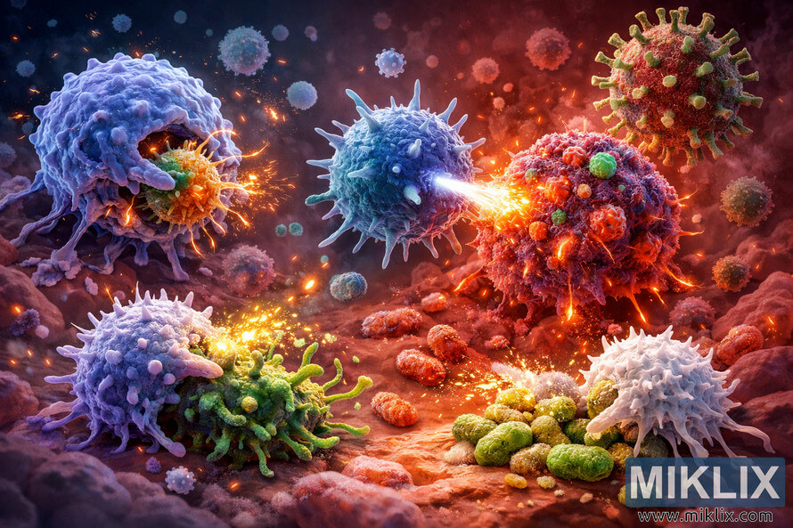
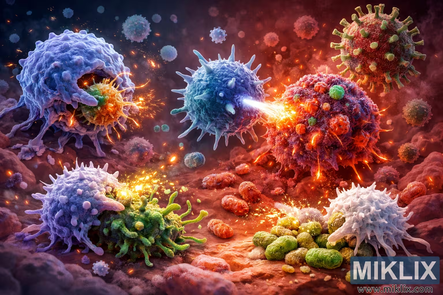
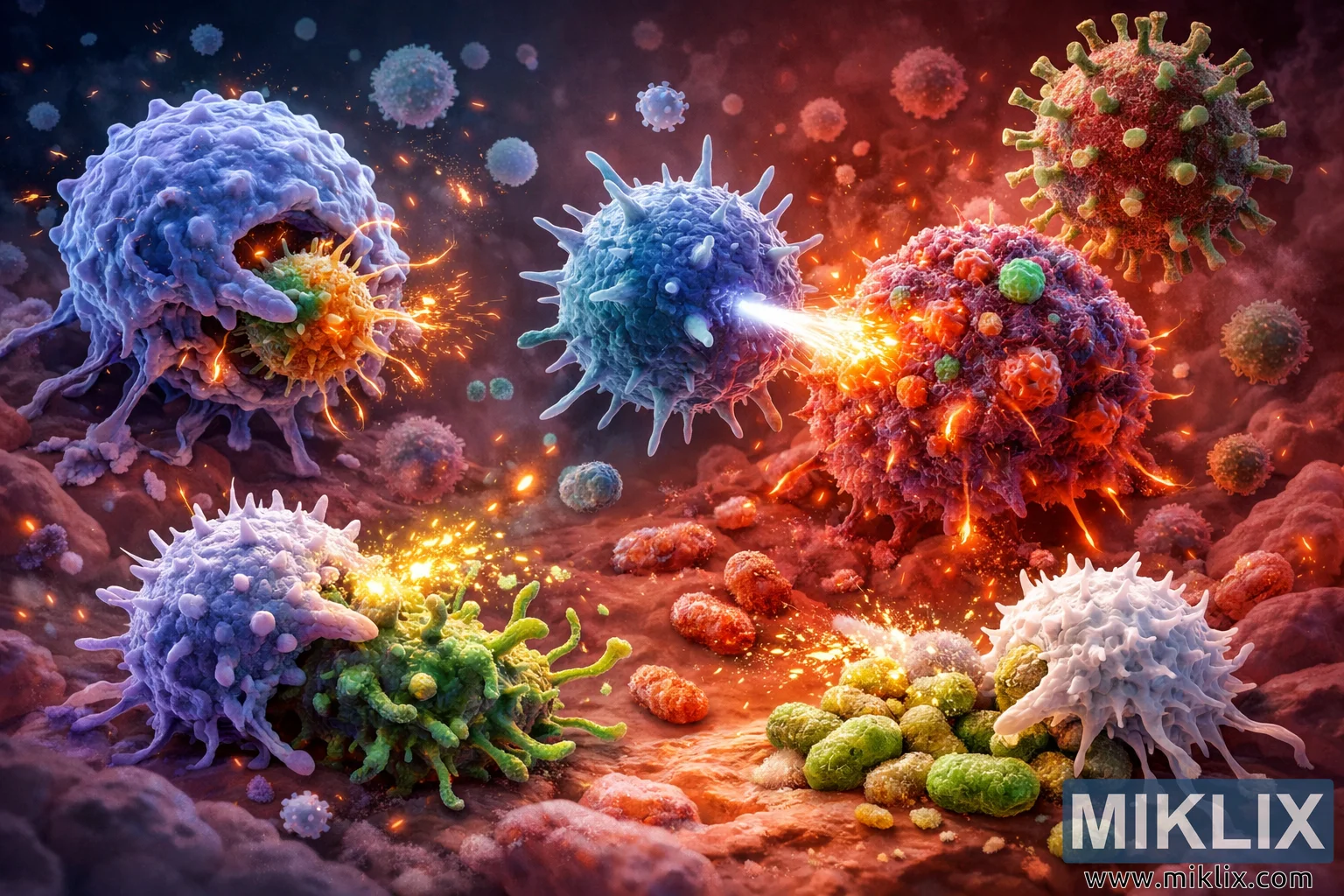
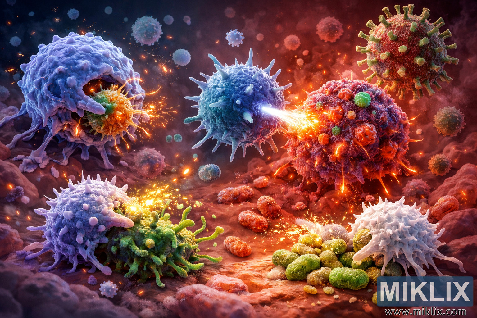
A detailed digital illustration of immune system cells fighting infection, featuring a macrophage, T cell, natural killer cell, and neutrophils actively attacking bacteria and virus-infected cells in a glowing microscopic environment. The image highlights the dynamic, coordinated immune response with dramatic lighting, vivid colors, and scientifically inspired visual elements.
Microscopic Battle: Immune Cells Fighting Infection
この画像の利用可能なバージョン
以下のダウンロード可能な画像ファイルは、帯域幅の消費を抑えるためにファイルサイズが最適化されている当ウェブサイトの記事やページに埋め込まれている画像よりも、圧縮率が低く、解像度が高く、その結果、品質が高くなっています。
レギュラーサイズ (1,536 x 1,024)
大きいサイズ (3,072 x 2,048)
非常に大きなサイズ (4,608 x 3,072)
特大サイズ (6,144 x 4,096)
非常に大きなサイズ (1,048,576 x 699,051)
- まだアップロード中だ。)
画像説明
A highly detailed, scientifically inspired digital illustration captures a dramatic microscopic battle inside the human body, where immune system cells are actively fighting off invading pathogens. The scene is set in a reddish-pink, glowing environment that suggests the interior of tissue or blood vessels, with a soft gradient that transitions from deeper reds and magentas to lighter pinks and subtle purples. This hazy backdrop creates a sense of depth and immersion, as if the viewer has been shrunk down to witness the immune response firsthand.
On the left side of the image, a large macrophage dominates the foreground. This macrophage is rendered as a bluish-purple, spherical cell with a rough, irregular surface, almost like a textured, semi-translucent orb. Its membrane appears slightly uneven, with small ridges and bumps that hint at its dynamic, flexible nature. Extending from the macrophage are several pseudopodia—elongated, arm-like protrusions that reach out toward a nearby bacterium. The bacterium is depicted as a spherical, spiky organism with orange and green coloration, its surface covered in short, sharp projections that give it a menacing appearance. As the macrophage engulfs the bacterium, glowing tendrils of light connect the two, visually representing the process of phagocytosis. These luminous strands suggest energy transfer and cellular activity, emphasizing the macrophage’s role as a frontline defender that consumes and neutralizes pathogens.
In the center of the composition, a powerful T cell serves as one of the main focal points. This T cell is a large, blue cell with spike-like protrusions radiating from its surface, giving it a starburst-like silhouette. The cell’s membrane is smooth yet studded with these protrusions, which symbolize receptors and activation structures. From the T cell, a bright beam of light extends toward an infected cell positioned slightly to the right. The infected cell is rendered in red and purple tones, with its surface covered in numerous virus particles. These viral structures cling to the cell’s membrane, and from them, fiery tendrils emerge as the cell undergoes destruction. The beam of light from the T cell appears almost like a focused energy ray, visually conveying the targeted attack that T cells perform when recognizing infected or abnormal cells. The interaction between the T cell and the infected cell is intense and dynamic, with glowing fragments and sparks suggesting the breakdown of the infected cell’s structure.
Below and slightly to the left of the central T cell, a natural killer (NK) cell adds another layer of action to the scene. The NK cell is depicted as a large, round, light purple cell covered in spiky protrusions, similar in some ways to the T cell but with a distinct coloration and texture. Its surface spikes give it a vigilant, aggressive appearance, emphasizing its role in identifying and destroying compromised cells. The NK cell is shown attacking a green, virus-infected cell that has tentacle-like protrusions extending outward. This infected cell appears distorted and irregular, with its tentacles reaching into the surrounding space as if trying to spread the infection further. From the NK cell, a burst of energy is released toward the infected cell, illustrated as a concentrated flare of light and particles. This visual effect highlights the NK cell’s ability to induce cell death in infected or cancerous cells, reinforcing the theme of active immune defense.
On the lower right side of the image, two white, spiky neutrophils are engaged in combat with a cluster of bacteria. The neutrophils are depicted as bright, white cells with numerous sharp protrusions, giving them a star-like shape. Their surfaces are slightly translucent, with subtle shading that suggests internal granules. Near these neutrophils is a group of greenish-yellow bacteria, clustered together in a small colony. One of the neutrophils is shown spewing enzymes and antimicrobial substances onto the bacteria, represented by a stream of glowing particles and fluid-like shapes that cascade over the bacterial cluster. This visual conveys the neutrophils’ role in releasing toxic substances to neutralize and digest pathogens, adding another layer of complexity to the immune response depicted.
Throughout the scene, small, round, glowing blue particles float in the environment. These particles resemble antibodies, tiny yet potent components of the immune system that bind to pathogens and mark them for destruction. Their soft glow and scattered distribution create a sense of pervasive immune activity, as if the entire space is saturated with defensive molecules. Interspersed among these antibodies are fiery particles and tendrils of light, which further emphasize the intensity of the ongoing battle. These glowing elements contribute to the dynamic lighting of the illustration, casting subtle reflections and highlights on the surfaces of cells and pathogens.
In the background, additional pathogens add depth and context to the scene. On the upper right, a large, spiky virus particle stands out, rendered in reddish tones with green spikes protruding from its surface. This virus appears ominous and threatening, looming behind the main action as a reminder of the constant presence of infectious agents. Smaller bacteria and viral particles are scattered throughout the background, partially blurred to maintain focus on the central immune cells while still conveying the complexity of the microscopic environment.
The lighting in the illustration is dramatic and dynamic, with bright highlights on the immune cells and pathogens, and softer, diffused light in the background. The interplay of cool and warm tones—blue and purple for immune cells, red, orange, and green for pathogens—creates strong visual contrast that helps distinguish defenders from invaders. The depth of field is moderate, keeping the main immune cells and their immediate targets in sharp focus while gently blurring more distant elements. This selective focus guides the viewer’s eye through the composition, from the macrophage on the left to the central T cell and NK cell, and finally to the neutrophils on the right.
Overall, the illustration presents a vivid, engaging visualization of the immune system in action. Each cell type is clearly differentiated by color, shape, and behavior, allowing viewers to appreciate the coordinated, multifaceted nature of the body’s defense mechanisms. The combination of scientific detail and artistic flair makes the image both educational and visually striking, capturing the complexity and intensity of the microscopic battle against infection.
画像は関連するものです: Health Benefits of Chives: Nature's Nutritional Powerhouse