
이미지: Advanced Laboratory Researching Dill-Derived Anti-Cancer Compounds
게시됨:
마지막으로 업데이트되었습니다: 2026년 7월 23일 오후 6시 43분 13초 UTC
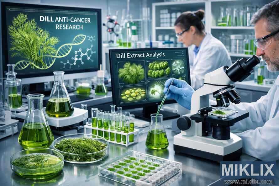
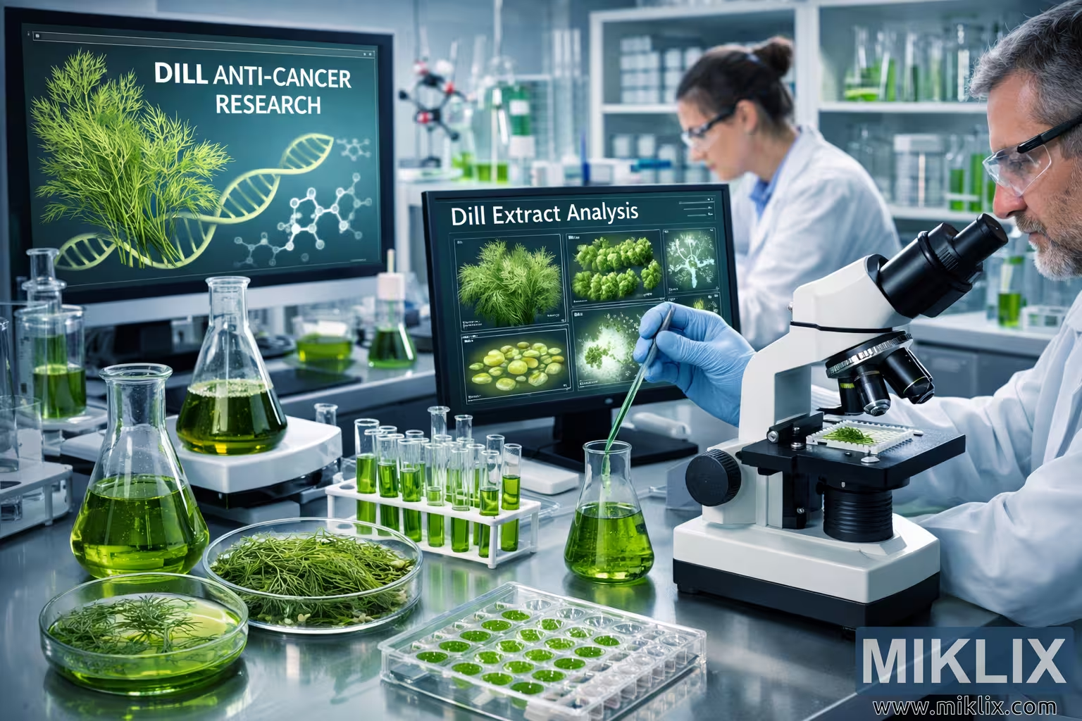
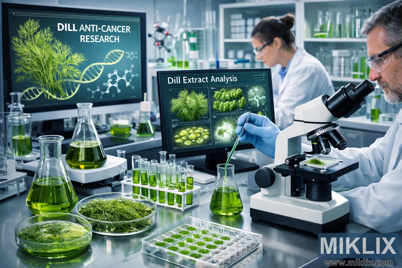
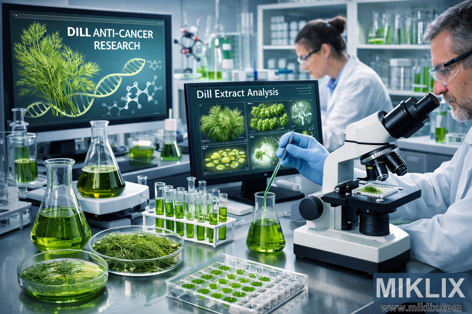
A detailed, high-resolution landscape image of a cutting-edge scientific laboratory researching anti-cancer compounds derived from dill. The scene shows scientists in lab coats and protective eyewear handling green dill extracts, pipetting samples, and analyzing data on a computer labeled "Dill Extract Analysis." Petri dishes with fresh dill, a compound microscope examining a dill sample, multi-well plates, test tubes, and flasks of green solutions fill the foreground, while the background displays beakers on a magnetic stirrer, a DNA helix model, and a large screen titled "DILL ANTI-CANCER RESEARCH." The cool, clean color palette and organized environment emphasize rigorous, modern research into the potential anti-cancer properties of dill.
Advanced Laboratory Researching Dill-Derived Anti-Cancer Compounds
이 이미지의 사용 가능한 버전
아래에서 다운로드할 수 있는 이미지 파일은 이 웹사이트의 기사 및 페이지에 삽입된 이미지보다 압축률이 낮고 해상도가 높으며, 대역폭 소비를 줄이기 위해 파일 크기에 더 최적화되어 있어 품질이 더 높습니다.
일반 사이즈 (1,536 x 1,024)
대형 사이즈 (3,072 x 2,048)
매우 큰 크기 (4,608 x 3,072)
초대형 사이즈 (6,144 x 4,096)
코믹하게 큰 사이즈 (1,048,576 x 699,051)
- 아직 업로드 중... ;-)
이미지 설명
The image depicts a modern scientific research laboratory dedicated to investigating anti-cancer compounds derived from dill. The scene is captured in a wide, landscape-oriented composition that allows the viewer to take in both the detailed activity on the laboratory bench and the broader environment of the research space. The overall atmosphere is clean, clinical, and highly organized, with a cool color palette dominated by whites, silvers, and various shades of green that visually emphasize the presence of dill extracts and related samples.
In the foreground on the left side, a glass laboratory bench serves as the primary surface for sample preparation. Resting on this bench is a petri dish filled with fresh dill sprigs, their feathery leaves and vibrant green color clearly visible. Next to it, another petri dish contains a green liquid representing dill extract, its smooth surface reflecting the laboratory lights. Behind these dishes stands a glass Erlenmeyer flask filled with a similar green solution, hinting at a larger volume of dill-derived compounds being prepared or stored for ongoing experiments.
On the right side of the foreground, a compound microscope is positioned prominently. Its objective lenses are angled downward, focused on a glass slide that holds a small sample of dill or dill-derived material. The microscope’s metallic body and precise mechanical components convey a sense of technical sophistication. The presence of the microscope underscores the microscopic level at which the research is conducted, suggesting that scientists are examining cellular responses, molecular structures, or other fine details related to the anti-cancer potential of dill.
At the center of the image, a scientist wearing a white lab coat, blue latex gloves, and protective eyewear is engaged in a careful pipetting procedure. The scientist holds a pipette in one hand and a test tube in the other, transferring a measured amount of green liquid—presumably dill extract—into the test tube. The posture and focus of the scientist convey concentration and precision, reflecting the meticulous nature of laboratory work. The blue gloves and clear safety glasses emphasize adherence to safety protocols and sterile technique.
To the right of this central figure, another scientist is seated or standing near a computer workstation. This scientist is also dressed in a white lab coat and protective eyewear, and is closely examining data displayed on a large monitor. The screen shows the heading "Dill Extract Analysis" along with visual elements such as molecular diagrams, images of cell cultures, a stylized DNA helix, and various charts and graphs. These graphical elements suggest that the research involves both experimental observation and computational analysis, integrating biological data with chemical and molecular information to understand how dill compounds might inhibit or affect cancer cells.
In the background, the laboratory extends further, revealing additional equipment and instruments. Several beakers containing green liquids are placed on a magnetic stirrer, which emits a soft glow, indicating active mixing or agitation of solutions. Nearby, a molecular model constructed from colored spheres and rods represents a chemical structure, possibly illustrating the molecular configuration of a key dill-derived compound or a related anti-cancer agent. Shelves and storage units hold various bottles, containers, and glassware, all neatly arranged to maintain an orderly and efficient workspace.
On the left side of the background, a large digital display or wall-mounted screen shows the text "DILL ANTI-CANCER RESEARCH" accompanied by an illustration of dill, a DNA helix, and a molecular structure. This visual reinforces the central theme of the laboratory’s work: exploring how compounds found in dill might contribute to cancer prevention or treatment. The combination of botanical imagery with molecular and genetic symbols highlights the intersection of natural products research, chemistry, and molecular biology.
The laboratory bench in the foreground contains additional items that further emphasize the depth and complexity of the research. A petri dish with green cells is visible, suggesting that cell cultures are being exposed to dill extracts to observe changes in growth, morphology, or viability. A multi-well plate holds multiple samples of green liquid, each well potentially representing a different concentration, formulation, or experimental condition. Nearby, a rack of test tubes filled with green solutions stands upright, ready for further analysis or processing. A conical flask containing green extract adds to the impression of an active, ongoing series of experiments.
To the right, a model of a DNA helix stands as a symbolic representation of genetic research and the molecular mechanisms underlying cancer. Its spiral form and colored segments draw the eye and connect the visual narrative of the image to the idea that dill-derived compounds may interact with cellular or genetic pathways. In the far background, more laboratory equipment, glassware, and storage shelves can be seen, reinforcing the sense that this is a fully equipped, professional research facility.
The lighting in the laboratory is bright and evenly distributed, creating a crisp, high-contrast environment where every detail is clearly visible. Reflections on glass surfaces, the shine of metal instruments, and the glow from electronic screens contribute to a high-tech ambiance. The dominant green tones of the dill samples and extracts stand out against the neutral whites and grays of the lab coats, walls, and equipment, visually signaling the central role of dill in the research.
Overall, the image conveys a narrative of scientific rigor and innovation. The scientists appear deeply engaged in their work, one focusing on the precise handling of dill extracts and the other analyzing complex data. The combination of traditional laboratory tools—such as microscopes, flasks, and petri dishes—with modern digital displays and molecular models suggests a multidisciplinary approach that spans chemistry, biology, and data science. The viewer is left with the impression that this laboratory is at the forefront of exploring how natural compounds from dill might contribute to the development of new anti-cancer strategies, all within a carefully controlled and thoughtfully designed research environment.
이 이미지는 다음과 관련이 있습니다: Health Benefits of Dill: Your Complete Guide to This Powerful Herb