
Gambar: Comparison of Healthy Retina Versus Damaged Retina
Diterbitkeun:
Panungtungan diropéa: 7 Séptémber 2026 jam 6.33.23 UTC
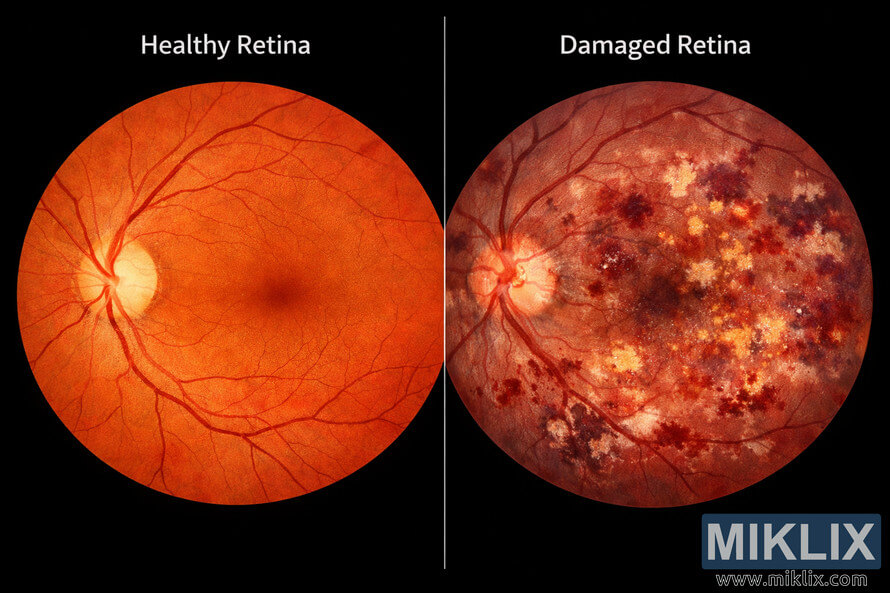
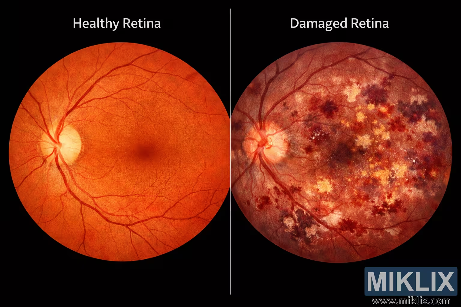
Detailed medical comparison image illustrating the differences between a healthy retina and a damaged retina, highlighting optic disc clarity, vessel integrity, and pathological changes.
Comparison of Healthy Retina Versus Damaged Retina
Versi anu sayogi tina gambar ieu
File gambar anu sayogi kanggo diunduh di handap ieu kirang dikomprés sareng résolusina langkung luhur - sareng akibatna, kualitasna langkung luhur - tibatan gambar anu dipasang dina artikel sareng halaman dina halaman wéb ieu, anu langkung dioptimalkeun pikeun ukuran file supados ngirangan konsumsi bandwidth.
Ukuran biasa (1,536 x 1,024)
Ukuran ageung (3,072 x 2,048)
Ukuran anu ageung pisan (4,608 x 3,072)
Ukuran anu ageung pisan (6,144 x 4,096)
Ukuran anu ageung pisan (1,048,576 x 699,051)
- Masih kénéh di upload... ;-)
Pedaran gambar
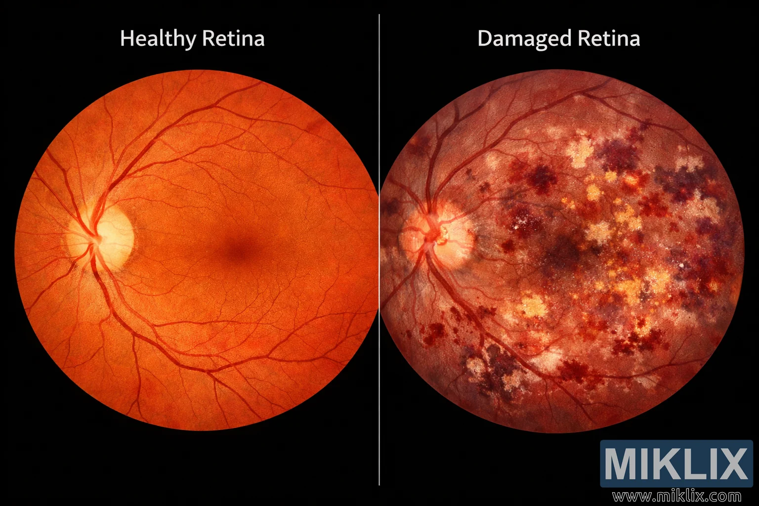
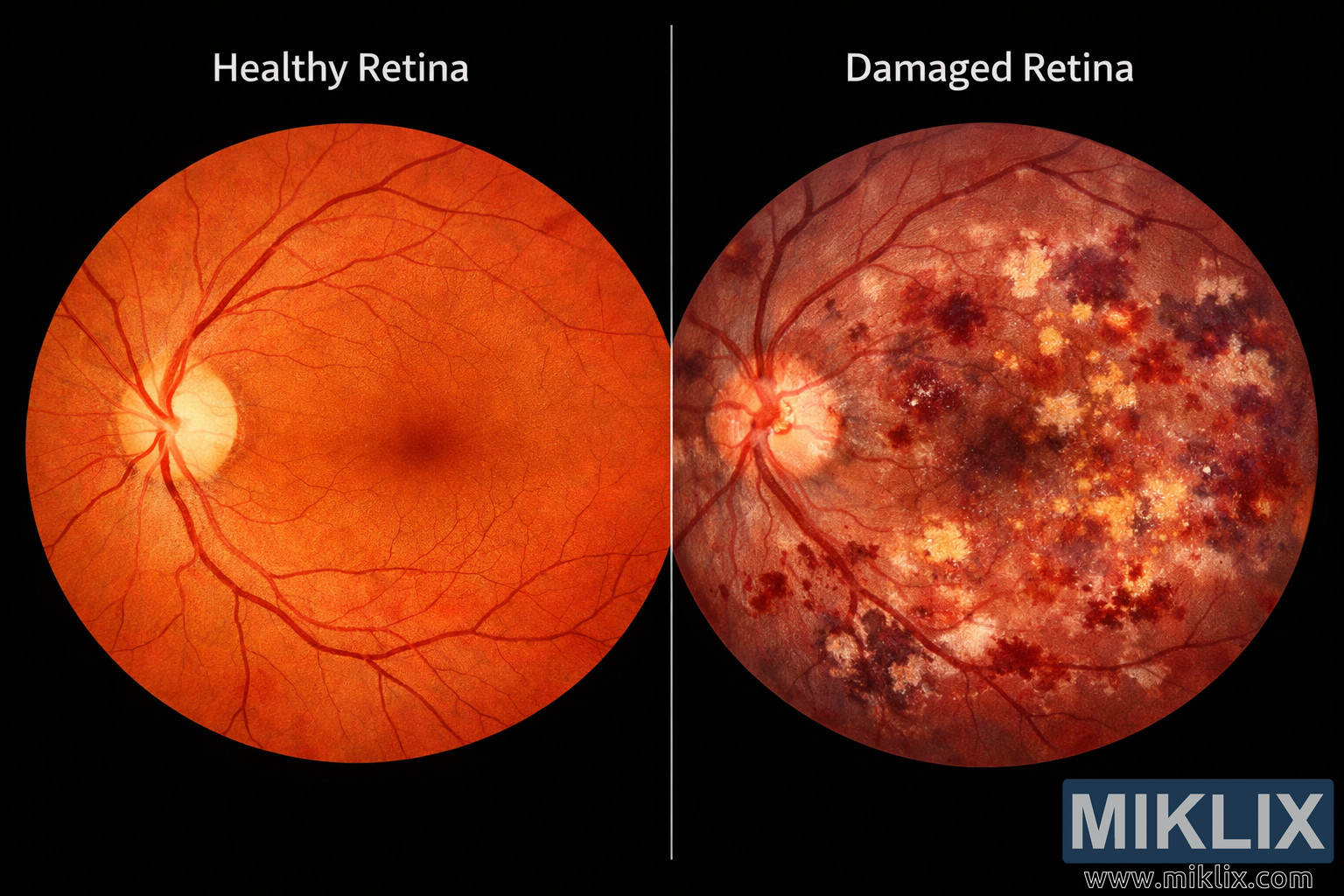
This image presents a detailed medical comparison between a healthy retina and a damaged retina, designed to visually communicate the structural and functional differences that occur due to retinal disease or injury. The composition is split into two halves by a vertical dividing line, creating a clear contrast between the left side, labeled 'Healthy Retina,' and the right side, labeled 'Damaged Retina.' The overall layout is symmetrical, with both retinas depicted in circular frames that mimic the natural curvature of the eye’s interior.
On the left side, the healthy retina is rendered in vibrant shades of orange and red, representing the rich vascularization and normal pigmentation of a well-functioning eye. The optic disc, located slightly off-center, appears as a pale yellowish-white circle where the optic nerve enters the retina. From this disc, fine blood vessels radiate outward in smooth, branching patterns, maintaining consistent thickness and coloration. The macula, positioned near the center, is visible as a darker, reddish spot that serves as the focal point for high-resolution vision. The overall texture of the healthy retina is uniform, with no visible lesions, hemorrhages, or discolorations. The lighting emphasizes clarity and vitality, suggesting optimal oxygenation and nutrient flow through the retinal tissue.
In contrast, the right side of the image depicts the damaged retina, characterized by a darker, reddish-brown hue and irregular patterns of light and shadow. The optic disc appears swollen and less defined, with blurred edges indicating inflammation or edema. The retinal surface is marred by multiple pathological features: dark red blotches representing hemorrhages, yellowish-white patches of exudates, and cloudy regions that suggest scarring or fluid accumulation. These visual cues correspond to common retinal disorders such as diabetic retinopathy, macular degeneration, or hypertensive retinopathy. The blood vessels in this section appear distorted—some are narrowed, others dilated, and several show signs of leakage. The overall impression is one of compromised structure and impaired function, with the damaged retina appearing congested and fragile compared to the healthy counterpart.
The central dividing line serves as both a visual and conceptual boundary, emphasizing the stark difference between health and disease. Above each half, the labels 'Healthy Retina' and 'Damaged Retina' are clearly printed in white, sans-serif text, ensuring immediate comprehension. The background is black, providing strong contrast that enhances the visibility of the retinal details and draws attention to the color variations. The lighting is even and clinical, avoiding artistic embellishment to maintain scientific accuracy.
This comparison image is valuable for educational and diagnostic purposes, helping viewers understand how retinal pathology manifests visually. It highlights key indicators such as optic disc clarity, vessel integrity, and the presence of hemorrhages or exudates. The healthy retina demonstrates the ideal state of ocular health—clear, well-structured, and vibrant—while the damaged retina reveals the consequences of disease progression, including tissue degradation and vascular instability.
Overall, the image conveys a powerful message about the importance of early detection and treatment in preserving vision. It serves as a visual guide for medical students, ophthalmologists, and patients alike, illustrating how subtle changes in retinal appearance can signify serious underlying conditions. The juxtaposition of the two retinas encapsulates the delicate balance between health and pathology within the human eye, reminding viewers of the intricate beauty and vulnerability of this essential organ.
Gambar aya hubunganana sareng: Pumpkins: The Nutrient-Packed Superfood You Should Eat Year-Round