
Image: Microscopic View of Ale Yeast Cells During Fermentation
Published: September 23, 2026 at 9:53:23 PM UTC
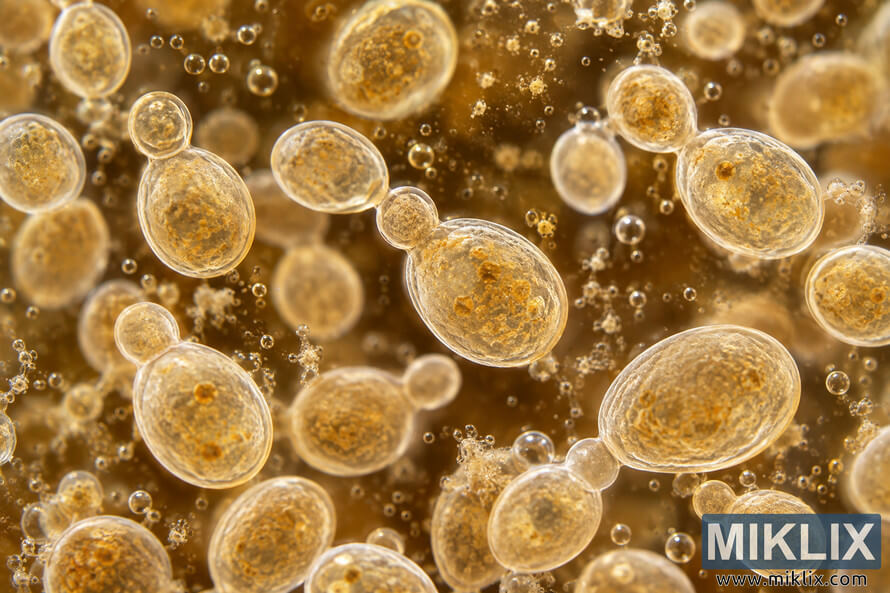
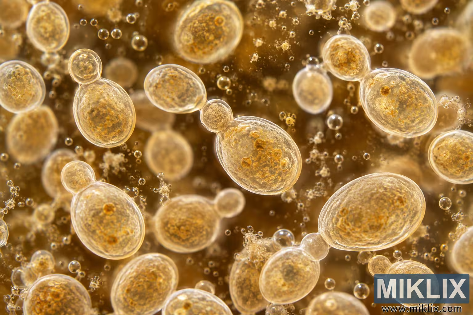
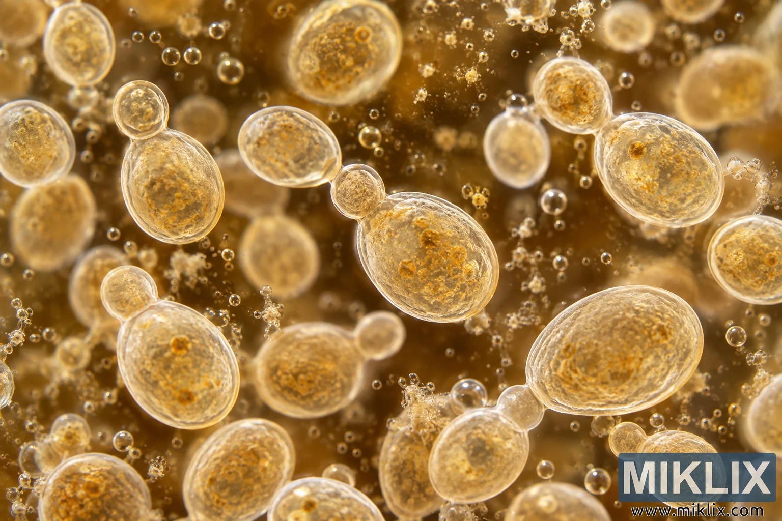
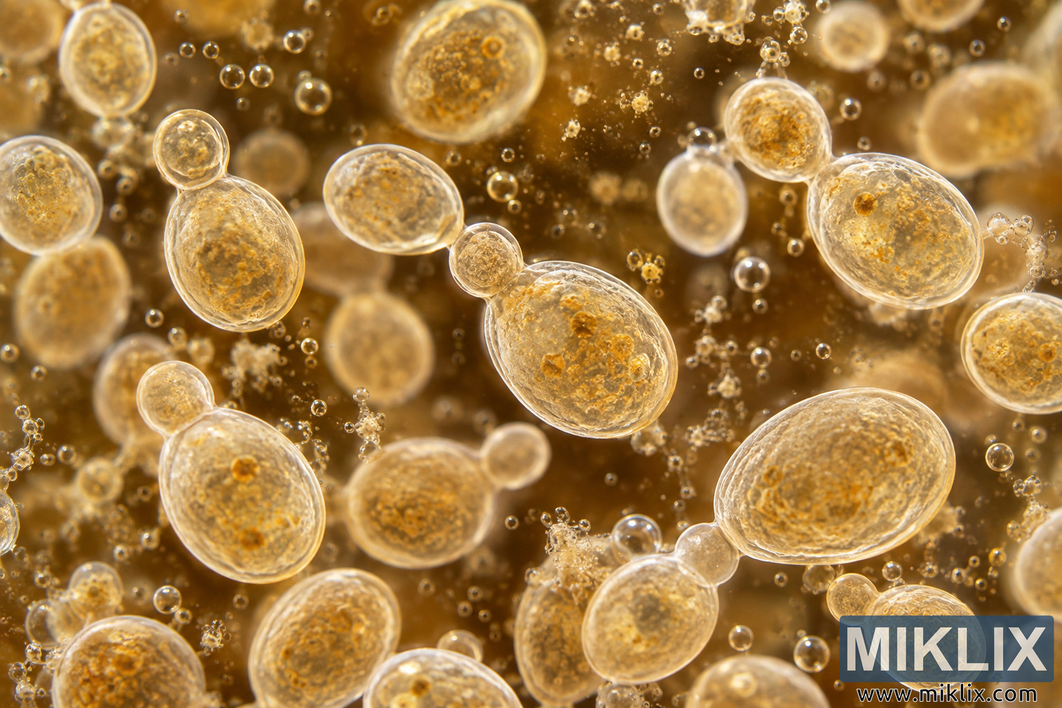
Detailed microscopic image of budding ale yeast cells during fermentation, suspended in warm amber liquid with tiny bubbles and textured cellular structures.
Available versions of this image
The image files available for download below are less compressed and higher resolution - and as a result of that, higher quality - than the images embedded in articles and pages on this website, which are more optimized for file size in order to reduce bandwidth consumption.
Regular size (1,536 x 1,024)
Large size (3,072 x 2,048)
Very large size (4,608 x 3,072)
Extra large size (6,144 x 4,096)
Comically large size (1,048,576 x 699,051)
- Still uploading... ;-)
Image description
A highly detailed, photorealistic microscopic close-up depicts ale yeast cells suspended in an amber-colored liquid during active fermentation. The landscape-oriented composition fills the entire frame with dozens of individual yeast cells at varying distances from the viewer, creating a strong impression of depth within a microscopic environment. The most prominent cells appear as translucent oval or rounded structures with clearly defined, luminous outer boundaries. Their interiors contain intricate granular textures in warm golden, honey, and pale brown tones, suggesting the complex internal material of living yeast cells. Several of the larger cells display smaller rounded protrusions attached to their surfaces, visually representing the budding process by which yeast cells reproduce. These budding pairs are distributed throughout the scene and provide an immediate visual indication that the microorganisms are biologically active.
The central area of the image contains one of the most visually prominent yeast cells. It is a large, nearly oval cell positioned slightly to the right of center, with a smaller spherical bud attached near its upper-left edge. The cell membrane is rendered as a bright, translucent rim that catches the surrounding light, while the interior has a dense, mottled appearance composed of fine golden particles and darker amber structures. Nearby cells show similar characteristics but differ subtly in size, orientation, internal texture, and stage of budding. This variation gives the microscopic scene a natural, organic quality rather than presenting the cells as uniform or diagrammatic objects.
On the left side of the frame, another large budding yeast cell is oriented vertically. A small, almost circular daughter cell is attached near its upper end, while the larger parent cell extends downward in an elongated oval form. Its outer boundary appears glasslike and softly illuminated. Toward the lower-left portion of the image, several additional cells occupy the foreground. These are rendered with relatively sharp detail and contain complex granular interiors. Other yeast cells in the middle distance and background gradually become softer and less distinct because of the shallow depth of field, reinforcing the impression that the viewer is looking through a three-dimensional suspension rather than at a flat scientific illustration.
The right half of the image contains several particularly large yeast cells. Near the upper-right area, a broad oval cell displays a bright translucent perimeter surrounding a textured golden interior. Farther down, an even larger oval cell dominates the lower-right quadrant. This cell is angled horizontally and has a smaller rounded structure attached close to one end, again suggesting budding. Fine internal patterns are visible through its semi-transparent surface. Smaller budding cells appear around it, including a compact pair near the bottom-right corner. Together, these organisms create a dense but balanced arrangement across the wide frame.
Surrounding the yeast cells are numerous tiny spherical bubbles and irregular suspended particles. The bubbles range from extremely small pinpoints to larger, clearly defined spheres with reflective highlights. Their presence evokes the production and movement of carbon dioxide associated with fermentation. Some bubbles occur individually, while others form loose clusters between the cells. Irregular pale fragments and granular particles are also dispersed throughout the liquid, adding complexity to the microscopic environment. These elements help communicate that the cells are suspended in an actively fermenting medium rather than isolated against a clean laboratory background.
The overall color palette is dominated by warm amber, gold, caramel, and honey-brown hues. The background liquid is substantially darker than the translucent cell membranes, producing strong visual separation between the microorganisms and their surroundings. Light appears to pass through or around the cells, creating glowing rims and subtle highlights that emphasize their rounded three-dimensional forms. The brightest edges approach a creamy off-white or pale gold, while deeper portions of the liquid shift toward rich brown and dark amber. This warm coloration visually connects the scientific subject with ale fermentation without showing any brewing equipment, packaging, logos, labels, or recognizable commercial brands.
Depth of field is an important visual characteristic of the image. Cells in the primary focal plane are crisp enough to reveal fine textures along their membranes and within their interiors, whereas cells positioned farther away become progressively blurred. Some background organisms appear only as soft golden ovals or circles. The same depth effect applies to the bubbles and suspended particles, with certain bubbles rendered as sharply defined transparent spheres and others dissolving into soft highlights. This selective focus gives the scene the appearance of a macro or microscopic photograph captured through specialized optical equipment.
The image combines scientific subject matter with cinematic photographic presentation. Although the cells are depicted in a visually polished and dramatic manner, key recognizable characteristics of yeast are emphasized, especially their generally oval shapes and the presence of attached buds. The abundance of cells, bubbles, and suspended matter conveys vigorous biological activity. There is no text anywhere in the frame and no human subject, laboratory instrument, container, bottle, brewery logo, product label, or other branded element. Instead, the entire composition remains focused on the microscopic world of ale yeast during fermentation. The result is an immersive close-up that visually communicates yeast growth, budding, suspension in liquid, and fermentation activity through a richly textured field of glowing microorganisms and tiny gas bubbles.
The image is related to: Fermenting Beer with White Labs WLP039 East Midlands Ale Yeast