
Image: Microscopic View of Budding Yeast Cells
Published: October 5, 2026 at 4:26:46 PM UTC
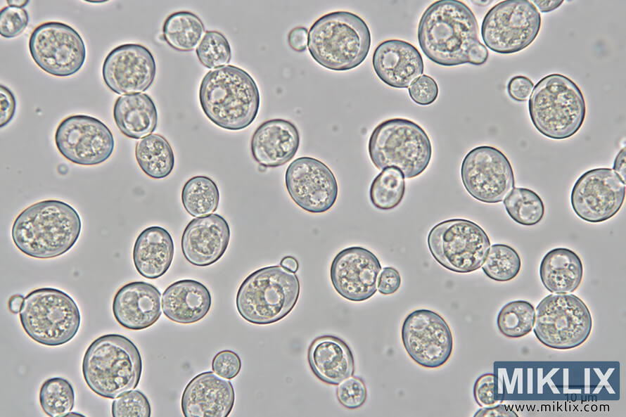
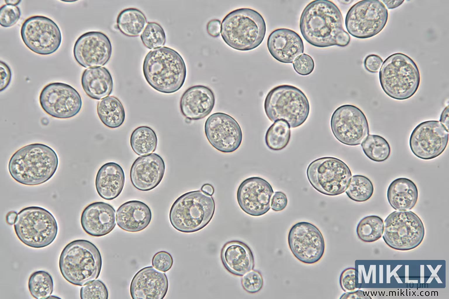
Scientific microscopy image showing numerous round and oval yeast-like cells, including budding forms, with visible internal detail and a 10 µm scale bar.
Available versions of this image
The image files available for download below are less compressed and higher resolution - and as a result of that, higher quality - than the images embedded in articles and pages on this website, which are more optimized for file size in order to reduce bandwidth consumption.
Regular size (1,536 x 1,024)
Large size (3,072 x 2,048)
Very large size (4,608 x 3,072)
Extra large size (6,144 x 4,096)
Comically large size (1,048,576 x 699,051)
- Still uploading... ;-)
Image description
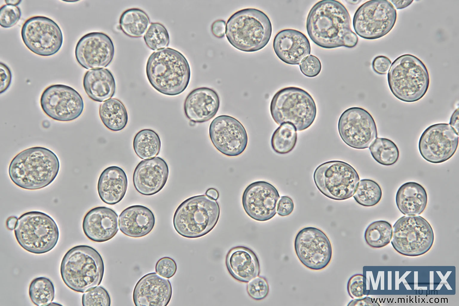
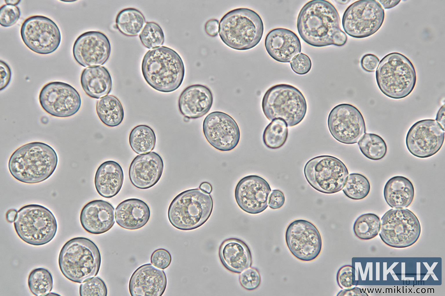
Landscape-oriented scientific microscopy image showing numerous round to oval yeast-like cells distributed across a pale gray field. The cells vary in size and are mostly separated from one another, although several occur as attached pairs or small groupings. Many larger cells have smaller rounded cells attached at their edges, visually consistent with budding morphology. Individual cells have dark, sharply defined outer boundaries with subtle colored fringes and translucent interiors containing irregular granular, mottled, or clustered structures. Several cells contain one or more prominent pale, nearly circular internal regions. The cells are scattered across the entire frame with substantial open background space between them. Some cells are partially cropped by the image edges, including cells along the top, left, and right margins. The background is light gray and softly textured, with faint small spots and blurred particulate-looking features. The overall appearance is consistent with a high-magnification brightfield or phase-contrast microscopy view, although the exact microscopy technique cannot be determined from the image alone. In the lower-right corner is a horizontal black scale bar with the readable label "10 µm" beneath it. No other text, labels, annotations, or branding are visible.
The image is related to: Fermenting Beer with White Labs WLP644 Saccharomyces brux-like Trois Yeast