
Image: Microscopic View of Yeast Cells During Fermentation
Published: October 9, 2026 at 10:23:12 PM UTC
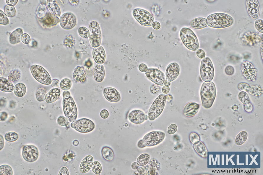
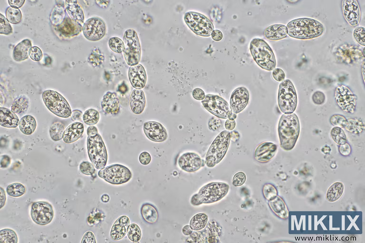
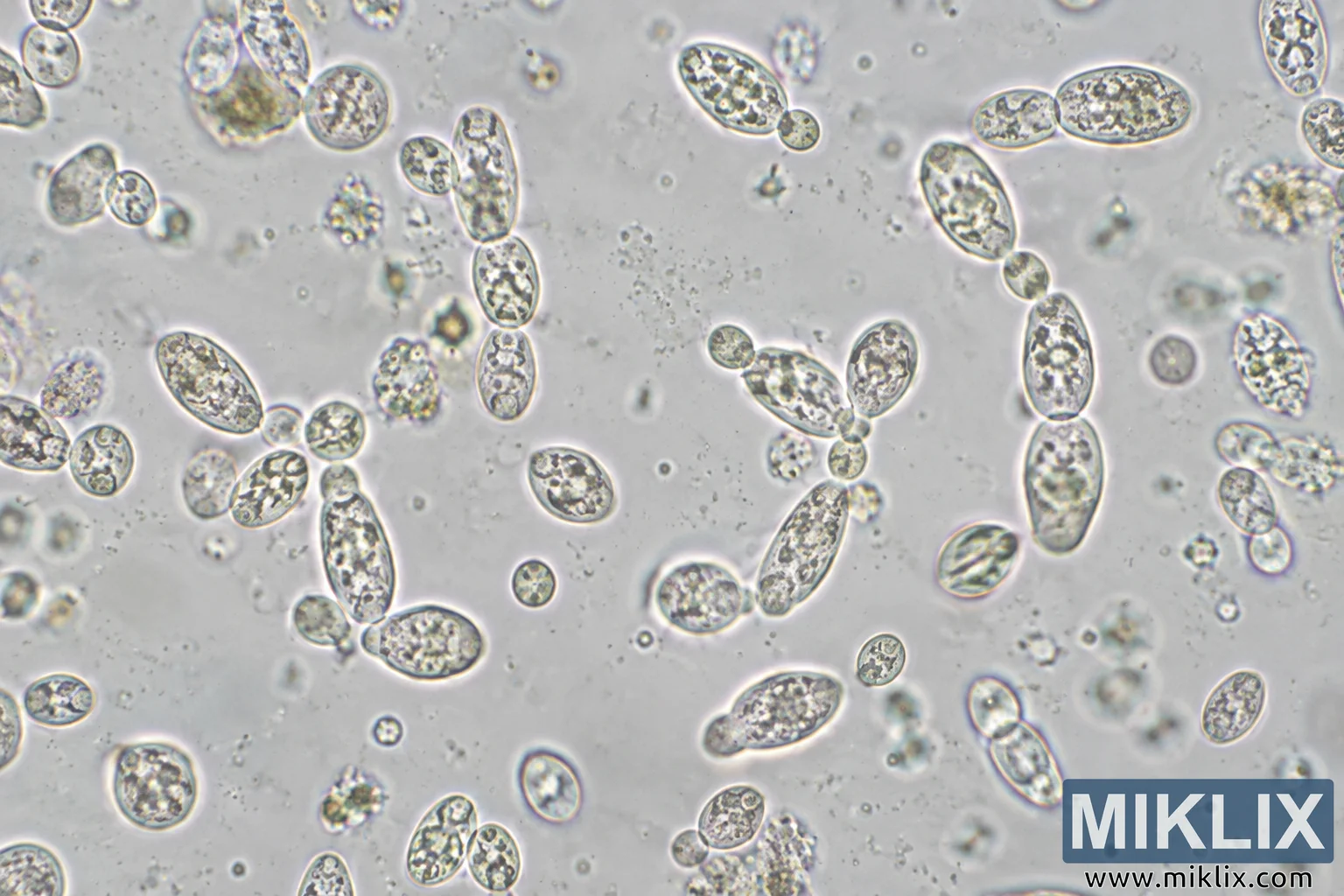
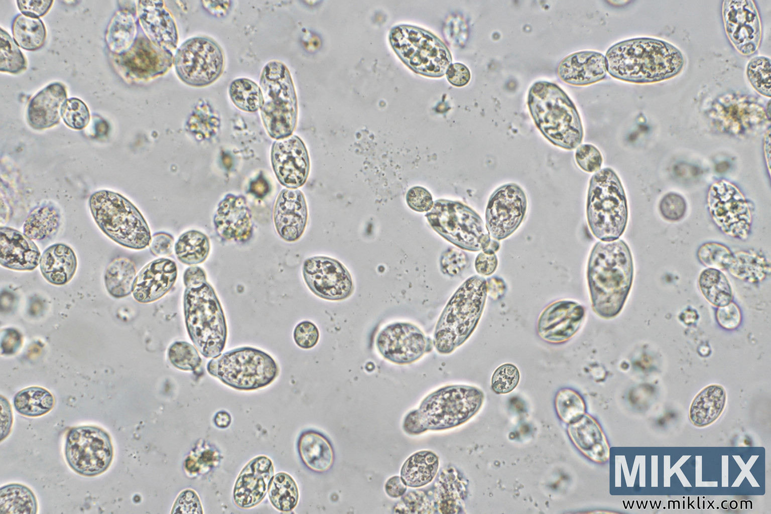
Detailed microscopic image of oval and elongated yeast-like cells with granular interiors, budding forms, small clusters, and scattered particles in a pale field.
Available versions of this image
The image files available for download below are less compressed and higher resolution - and as a result of that, higher quality - than the images embedded in articles and pages on this website, which are more optimized for file size in order to reduce bandwidth consumption.
Regular size (1,536 x 1,024)
Large size (3,072 x 2,048)
Very large size (4,608 x 3,072)
Extra large size (6,144 x 4,096)
Comically large size (1,048,576 x 699,051)
- Still uploading... ;-)
Image description
A landscape-format photomicrograph showing numerous individual and clustered yeast-like cells distributed across a pale gray, slightly mottled microscopic field. The cells are predominantly oval to elongated, with some nearly round forms. Many have bright, sharply defined outer edges surrounded by subtle halos, while their interiors contain irregular granular and mottled structures in gray, beige, muted yellow-green, and dark brown-gray tones. Several cells appear connected in pairs or short chains, and some smaller rounded cells are attached to larger oval cells, consistent visually with budding morphology, although the exact biological process cannot be confirmed from the image alone. Cell sizes and orientations vary throughout the frame. The highest concentration of clearly focused cells occurs across the left, central, and right portions of the image, while other cellular forms and particulate material are softer or blurred, suggesting different focal depths. Small circular particles, irregular specks, and diffuse translucent structures are scattered through the surrounding field. In the upper right area, two elongated oval cells lie horizontally next to each other, while below them a roughly vertical sequence of larger oval cells extends toward the center-right. Near the center, several oval cells and smaller rounded forms form a loose cluster. The lower half contains additional isolated cells and small groups, including prominent oval forms with granular interiors. The background is predominantly light gray to off-white with subtle variations in brightness. The overall appearance resembles phase-contrast or a similar light-microscopy technique, but the specific imaging method cannot be determined with certainty from the image alone. No laboratory equipment, scale bar, labels, annotations, logos, brands, or readable text are visible. The image itself does not provide sufficient information to independently verify the species identity of the depicted microorganisms.
The image is related to: Fermenting Beer with White Labs WLP645 Brettanomyces Claussenii Yeast