
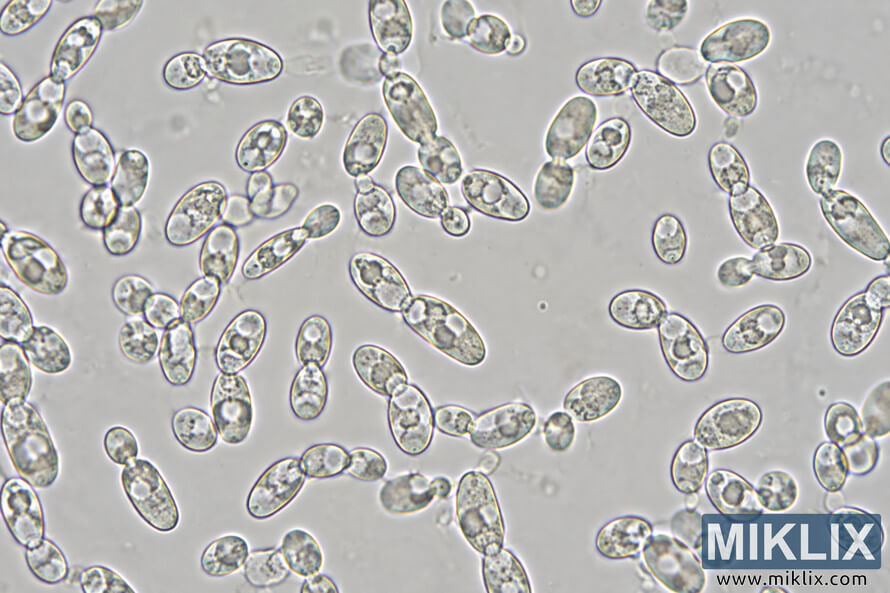
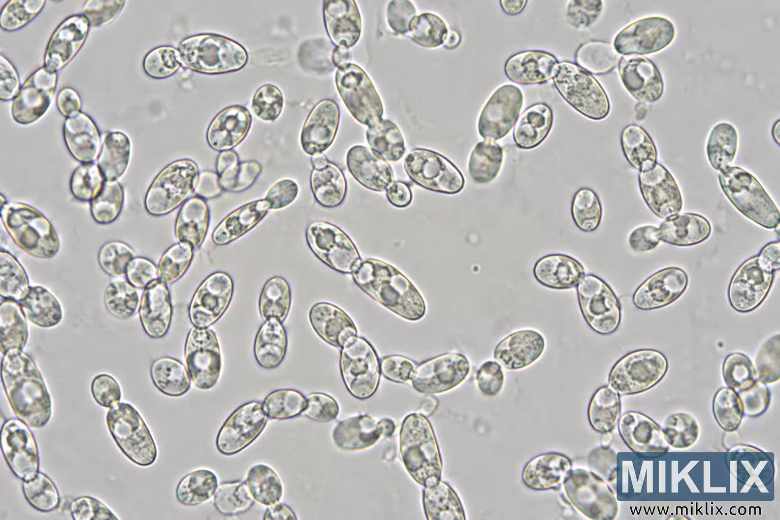
Image: Microscopic View of Brettanomyces bruxellensis Yeast Cells
Published: November 4, 2026 at 7:45:17 PM UTC
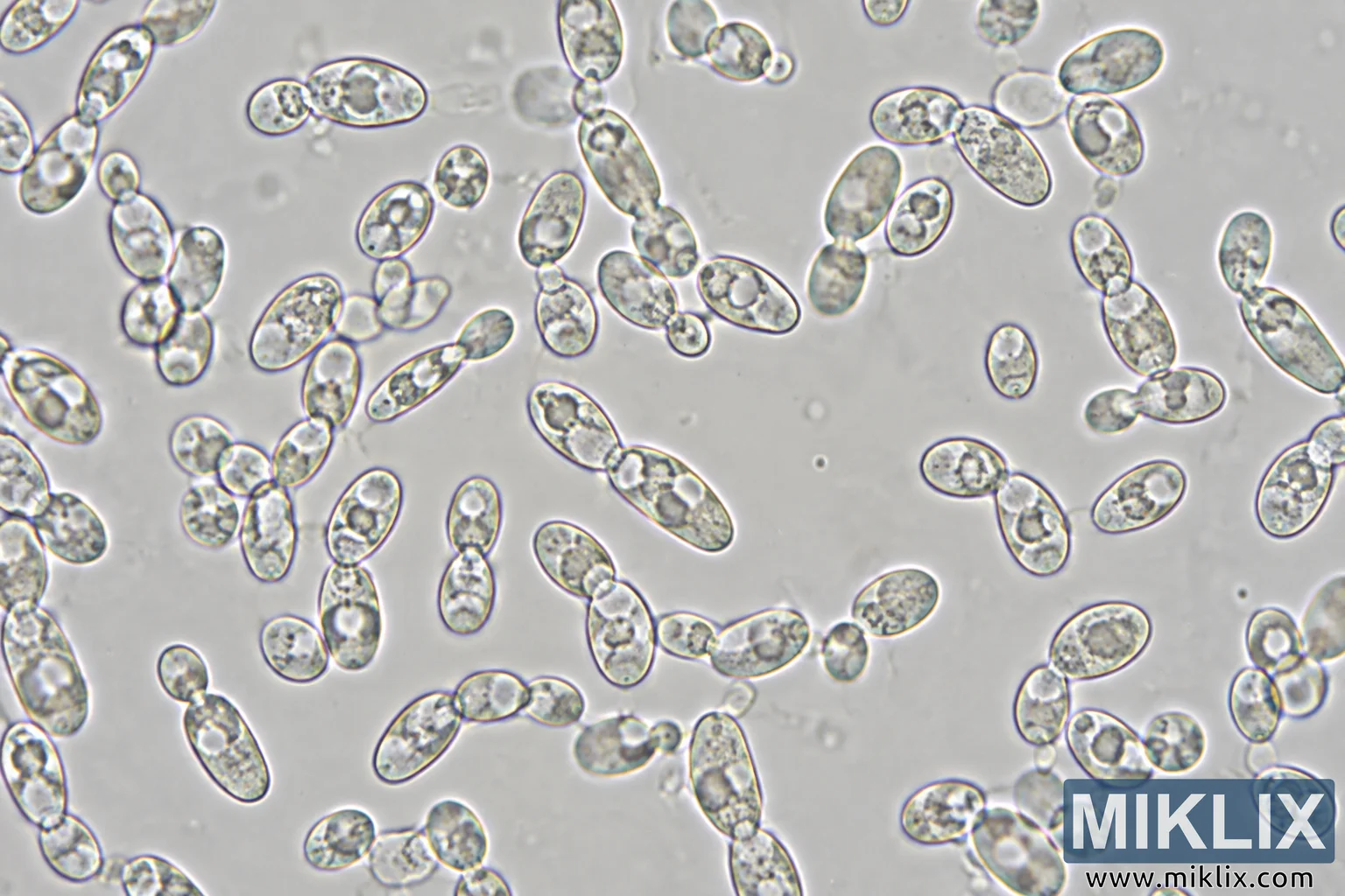
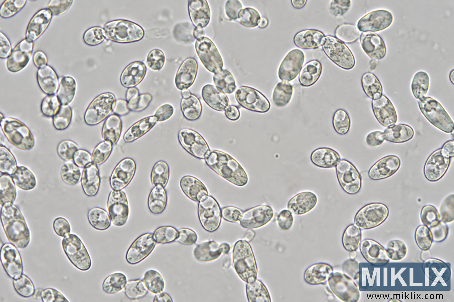
High-magnification microscopic image depicting Brettanomyces bruxellensis yeast-like cells in varied oval and elongated forms, with budding cells and small clusters.
Available versions of this image
The image files available for download below are less compressed and higher resolution - and as a result of that, higher quality - than the images embedded in articles and pages on this website, which are more optimized for file size in order to reduce bandwidth consumption.
Regular size (1,536 x 1,024)
Large size (3,072 x 2,048)
Very large size (4,608 x 3,072)
Extra large size (6,144 x 4,096)
Comically large size (1,048,576 x 699,051)
- Still uploading... ;-)
Image description
Landscape-oriented microscopic image showing numerous translucent, yeast-like cells distributed across a pale gray-white field. The cells are predominantly oval, elongated oval, teardrop-shaped, or slightly irregular, with substantial variation in size and shape. Many occur individually, while others appear as attached pairs, budding forms, small clusters, or short chains. Individual cells have clearly defined dark outlines with subtle blue-violet, yellow, and greenish color fringes along some edges, consistent with the visible optical characteristics of the microscopy image. Their interiors are mottled and granular, containing varying combinations of gray, beige, pale yellow, and nearly transparent regions. Many cells contain one or more rounded, relatively clear internal areas resembling vacuole-like structures, although their exact biological identity cannot be determined from the image alone. Several attached cells have narrow constrictions at their junctions, and cells in the short chains generally connect end-to-end. The field is moderately dense but contains an open, comparatively cell-free region near the center. Larger concentrations of cells occur toward the left side, along the upper portion, and in the lower-right quadrant. Some cells extend beyond the image boundaries, indicating that the visible frame represents only part of a larger microscopic field. The background is softly textured and slightly uneven in brightness, with faint blurred spots, small circular artifacts, and indistinct material visible between the sharply focused cells. Depth of field varies slightly: most prominent cells are sharply outlined, while some background features and a few cells or cell portions are softer. There are no macroscopic objects, labels, scale bars, annotations, logos, brands, or readable text. The image visually depicts yeast cells and was generated to represent Brettanomyces bruxellensis, but species identity cannot be independently confirmed from visual appearance alone.
The image is related to: Fermenting Beer with Wyeast 5112 Brettanomyces Bruxellensis Yeast