
Image: Microscopic View of Yeast Cells
Published: November 17, 2026 at 7:20:34 PM UTC
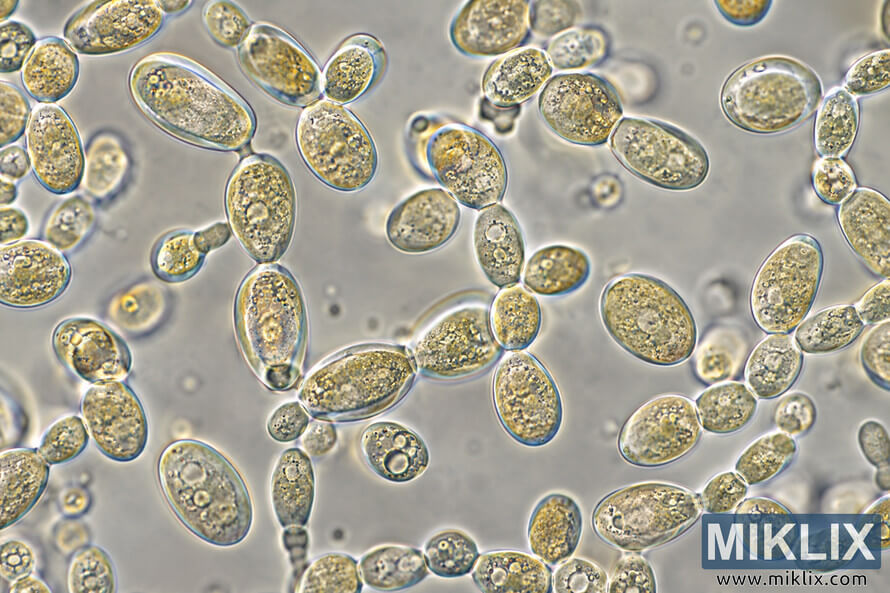
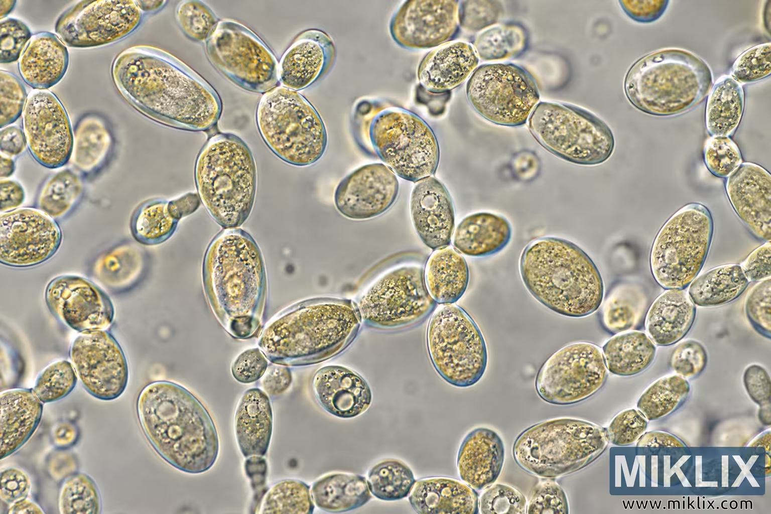
High-magnification microscopic image showing numerous oval and elongated yeast-like cells with granular interiors, budding forms, and translucent edges.
Available versions of this image
The image files available for download below are less compressed and higher resolution - and as a result of that, higher quality - than the images embedded in articles and pages on this website, which are more optimized for file size in order to reduce bandwidth consumption.
Regular size (1,536 x 1,024)
Large size (3,072 x 2,048)
Very large size (4,608 x 3,072)
Extra large size (6,144 x 4,096)
Comically large size (1,048,576 x 699,051)
- Still uploading... ;-)
Image description
A landscape-format microscopic image showing numerous oval to elongated, translucent cell-like structures distributed across a pale gray-beige background. The cells vary noticeably in size and orientation. Many appear individually, while others occur in attached pairs, short chains, or small clusters. Several structures have smaller rounded or oval bodies connected to larger cells, producing budding-like forms. The cell boundaries are sharply defined by bright refractive outlines with subtle blue-green and warm yellow edge coloration. Their interiors contain irregular granular textures, small dark specks, pale circular or oval inclusions, and occasional larger clear, vacuole-like areas. Some cells have comparatively smooth interiors, while others are densely granular. The structures are concentrated around the left, upper, central, and right portions of the frame, with a relatively open area of background near the upper-middle region. A prominent branching group of connected cells extends vertically through the center, while additional chains and clusters appear along the right side and lower edge. Objects outside the primary focal plane appear softer and more blurred, creating visible depth within the microscopic field. The background is softly mottled and contains faint circular and irregular out-of-focus features. Illumination gives many cell interiors a pale golden or amber tone against the neutral background. No scale bar, labels, symbols, branding, or other readable text are visible. The image visually resembles a high-magnification light-microscopy view of yeast cells; however, the specific organism or strain cannot be independently determined from visual appearance alone.
The image is related to: Fermenting Beer with Wyeast 5526 Brettanomyces Lambicus Yeast