
Imatge: Detailed Cross-Section of Human Skin Layers and Collagen Structure
Publicat:
Última actualització: 3 de setembre del 2026, a les 20:27:14 UTC
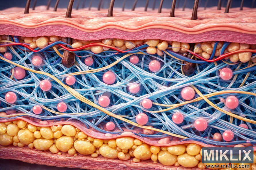
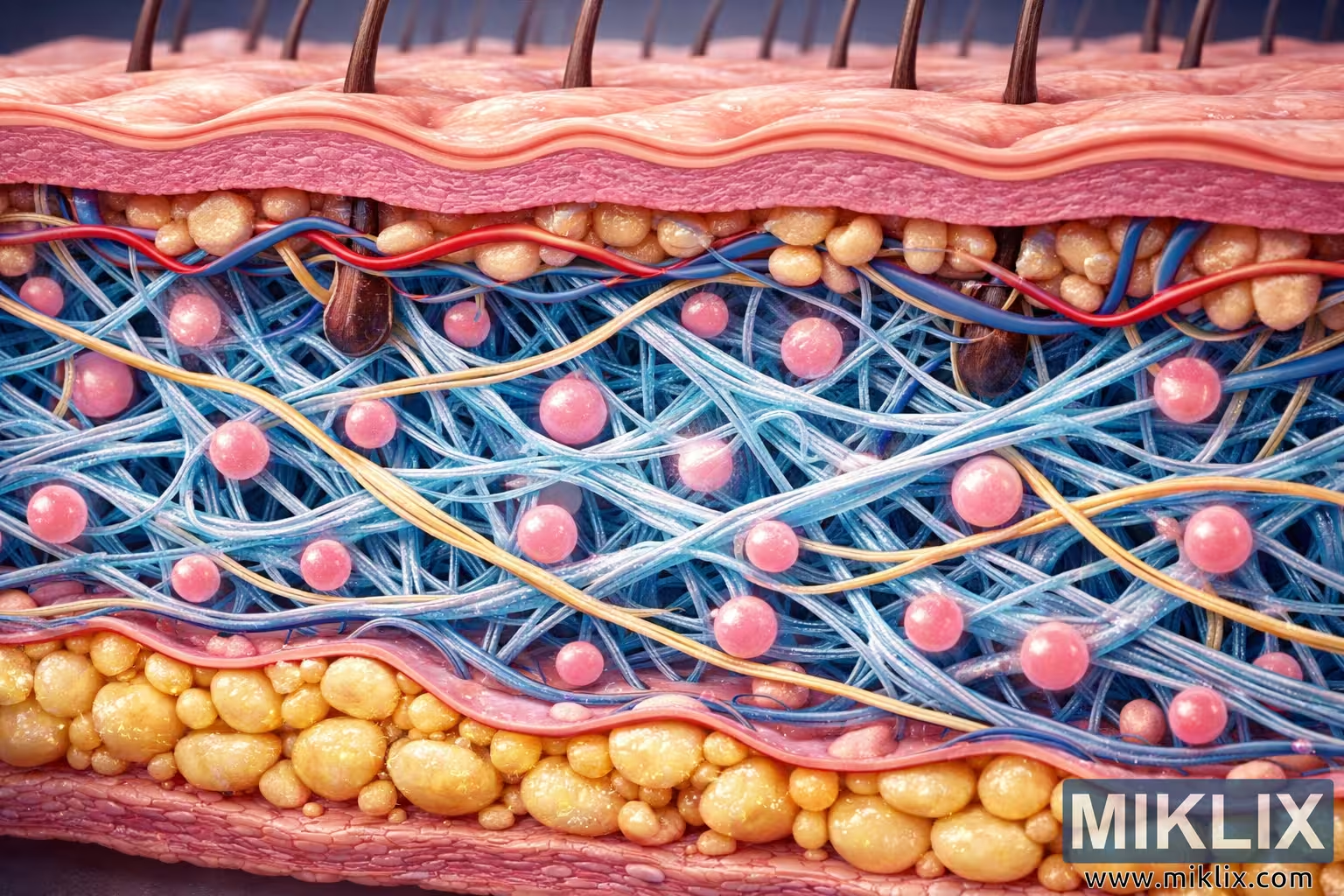
A high-resolution 3D illustration of human skin layers highlighting the collagen structure within the dermis. The image shows the epidermis, dermis, collagen fibers, fibroblasts, and subcutaneous fat cells in vivid detail.
Detailed Cross-Section of Human Skin Layers and Collagen Structure
Versions disponibles d'aquesta imatge
Els fitxers d'imatge disponibles per descarregar a continuació estan menys comprimits i tenen una resolució més alta (i, com a resultat, una qualitat més alta) que les imatges incrustades en articles i pàgines d'aquest lloc web, que estan més optimitzades per a la mida del fitxer per tal de reduir el consum d'ample de banda.
Mida normal (1,536 x 1,024)
Mida gran (3,072 x 2,048)
Mida molt gran (4,608 x 3,072)
Mida extra gran (6,144 x 4,096)
Mida còmicament gran (1,048,576 x 699,051)
- Encara s'està carregant... ;-)
Descripció de la imatge
This image presents a scientifically accurate and visually compelling 3D cross-sectional view of human skin, emphasizing the intricate collagen structure within the dermis. The composition is arranged horizontally in landscape orientation, allowing each layer of the skin to be clearly distinguished and studied in relation to the others. The topmost layer, the epidermis, is rendered in soft pink tones with subtle gradients that transition from light pink at the surface to deeper hues below. The surface of the epidermis is slightly undulating, representing the natural texture of the stratum corneum, which acts as a protective barrier. Tiny ridges and valleys are visible, giving the layer a realistic tactile quality.
Beneath the epidermis lies the dermis, the central focus of the image. This layer is depicted with remarkable detail, showing a dense network of collagen fibers intertwined with elastic strands. The collagen fibers appear as wavy, blue-white bands that crisscross in multiple directions, forming a resilient lattice that provides structural support to the skin. Between these fibers are small, glowing pink spheres representing fibroblasts—the cells responsible for producing collagen and maintaining the extracellular matrix. The fibroblasts are scattered throughout the dermis, creating a dynamic sense of biological activity.
Blood vessels are also visible within the dermis, portrayed as thin red and blue tubes weaving through the collagen network. These vessels symbolize the arteries and veins that supply oxygen and nutrients to the skin while removing waste products. Hair follicles extend downward from the epidermis into the dermis, each containing a dark brown hair shaft surrounded by sebaceous glands. The glands are shown as small, bulbous structures attached to the follicles, emphasizing their role in secreting oils that lubricate the skin and hair. Sweat glands are also present, depicted as coiled tubular structures that wind through the dermal layer, highlighting the skin’s thermoregulatory function.
The collagen matrix itself is the visual centerpiece of the image. The fibers are arranged in a semi-organized pattern, with varying thickness and curvature to reflect the natural complexity of connective tissue. The interplay of light and shadow across these fibers creates a sense of depth and dimensionality, making the collagen appear almost tangible. The blue-white coloration contrasts beautifully with the warmer tones of the surrounding tissue, drawing the viewer’s attention to the structural integrity that collagen provides.
Below the dermis lies the subcutaneous layer, also known as the hypodermis. This layer is composed primarily of adipose tissue, represented by clusters of rounded, yellow fat cells. Each cell is rendered with a glossy surface and subtle shading to convey volume and density. The fat cells are tightly packed, forming a cushion that insulates the body and absorbs mechanical shocks. Small blood vessels weave through this layer as well, maintaining the continuity of the circulatory system throughout the skin’s depth.
The lighting in the image is even and well-balanced, illuminating each layer without overpowering the natural color variations. The shadows cast by the collagen fibers and fibroblasts add realism and emphasize the three-dimensional structure. The overall color palette is harmonious, combining warm pinks and yellows with cool blues and whites to create a visually appealing and scientifically informative representation.
This illustration serves both educational and aesthetic purposes. It provides a clear understanding of how the skin’s layers interact and how collagen contributes to elasticity, strength, and youthful appearance. The meticulous attention to anatomical accuracy makes it suitable for medical education, dermatological research, and cosmetic science. The image captures the complexity of human skin as a living, dynamic organ—one that protects, senses, and regenerates.
In summary, this high-resolution depiction of skin layers and collagen structure offers a comprehensive visual study of the epidermis, dermis, and hypodermis. It highlights the collagen matrix as the foundation of skin integrity, supported by fibroblasts, blood vessels, glands, and fat cells. The result is a stunning fusion of art and science that reveals the hidden architecture beneath the surface of human skin.
La imatge està relacionada amb: Loquat: The Sweet Fruit Packed with Real Health Benefits