
Image: Dogwood Anthracnose Symptoms: Leaf Spots and Twig Dieback
Published: December 10, 2025 at 9:35:15 PM UTC
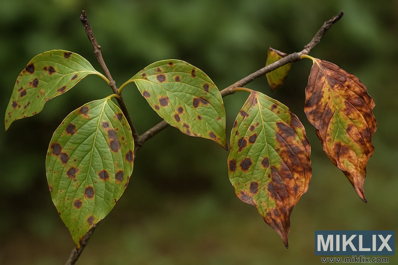
Detailed photograph of dogwood anthracnose symptoms, displaying dark brown leaf spots and twig dieback on a green branch.

Available versions of this image
The image files available for download below are less compressed and higher resolution - and as a result of that, higher quality - than the images embedded in articles and pages on this website, which are more optimized for file size in order to reduce bandwidth consumption.
Regular size (1,536 x 1,024)
Large size (3,072 x 2,048)
Very large size (4,608 x 3,072)
Extra large size (6,144 x 4,096)
Comically large size (1,048,576 x 699,051)
- Still uploading... ;-)
Image description
The image presents a high-resolution, landscape-oriented close-up of a dogwood branch affected by anthracnose, a fungal disease caused primarily by Discula destructiva. The composition centers on several elliptical dogwood leaves arranged along a thin, woody twig. The leaves are primarily green but exhibit widespread signs of infection in the form of irregularly shaped, dark brown to purplish lesions scattered across their surfaces. Each leaf shows varying degrees of damage: some have small, isolated spots, while others display extensive necrosis that causes browning and curling around the edges. The diseased tissue appears sunken and brittle, contrasting sharply with the still-living green portions of the leaves.
The twig itself reveals early signs of dieback, visible through a darkened, slightly shriveled tip where tissue death has begun to progress. Small cracks are visible along portions of the bark, suggesting that the fungus has invaded the vascular tissue beneath. These visual cues are characteristic of advanced anthracnose infections, in which both foliage and young stems are compromised, often leading to defoliation or branch death.
The image’s background is softly blurred, with an even, natural green tone that draws focus toward the sharply detailed leaves and twig in the foreground. This shallow depth of field emphasizes the contrast between healthy and diseased plant material, making the spots and discoloration highly visible. The lighting is diffuse and natural, avoiding harsh highlights while revealing subtle textures of leaf veins and epidermal tissue. The veins remain clearly defined in some of the less-damaged leaves, running in their typical arcuate pattern toward the leaf tip. In the more affected leaves, however, the venation is partially obscured by fungal lesions and spreading necrosis.
The overall color palette transitions from fresh green to deep brown, orange, and blackish tones, mirroring the typical progression of anthracnose damage as the fungus disrupts chlorophyll and induces cellular collapse. This vivid gradient visually narrates the disease’s destructive path from infection points to tissue death. A faint yellow halo is visible around some lesions, suggesting active fungal growth and toxin production at lesion margins.
From a diagnostic perspective, this image effectively illustrates both the foliar and twig symptoms used to identify dogwood anthracnose in the field. The pattern of spotting—typically more pronounced on shaded or lower leaves—and the dieback at twig tips are key indicators. The clarity and realism of the photo make it suitable for use in plant pathology guides, extension publications, and educational materials focused on disease recognition and forest health management.
The image is related to: A Guide to the Best Varieties of Dogwood Trees for Your Garden